US20060134728A1 - Microbial detection and quantification - Google Patents
Microbial detection and quantification Download PDFInfo
- Publication number
- US20060134728A1 US20060134728A1 US11/117,651 US11765105A US2006134728A1 US 20060134728 A1 US20060134728 A1 US 20060134728A1 US 11765105 A US11765105 A US 11765105A US 2006134728 A1 US2006134728 A1 US 2006134728A1
- Authority
- US
- United States
- Prior art keywords
- dye
- color
- control
- test
- dyes
- Prior art date
- Legal status (The legal status is an assumption and is not a legal conclusion. Google has not performed a legal analysis and makes no representation as to the accuracy of the status listed.)
- Granted
Links
- 0 */[NH+]=C\C=C\C=C/O.*N/C=C/C=C/C=O Chemical compound */[NH+]=C\C=C\C=C/O.*N/C=C/C=C/C=O 0.000 description 8
- PQHYSFLSXWWVBS-SNAWJCMRSA-N C=[N+](C)/C=C/C Chemical compound C=[N+](C)/C=C/C PQHYSFLSXWWVBS-SNAWJCMRSA-N 0.000 description 2
- YAGIKUGDXINHLL-ZPZFBZIMSA-L CC(=O)NC1=CC(S(=O)(=O)O[Na])=C(/N=N/C2=C3C(O)=CC(S(=O)(=O)O[Na])=CC3=CC=C2N)C=C1 Chemical compound CC(=O)NC1=CC(S(=O)(=O)O[Na])=C(/N=N/C2=C3C(O)=CC(S(=O)(=O)O[Na])=CC3=CC=C2N)C=C1 YAGIKUGDXINHLL-ZPZFBZIMSA-L 0.000 description 1
- HALVNEXTAQIUIP-UHFFFAOYSA-N CC1=C(C2=CC=C(C(C)(C)C)C=C2)C=C([N+]2=C(C3=CC(C(C)(C)C)=CC(C(C)(C)C)=C3)C=C(C3=CC=C(C(C)(C)C)C=C3)C=C2C2=CC(C(C)(C)C)=CC(C(C)(C)C)=C2)C=C1C1=CC=C(C(C)(C)C)C=C1.CC1=C(C2=CC=C(C(C)(C)C)C=C2)C=C([N+]2=C(C3=CC=C(C45CC6CC(CC(C6)C4)C5)C=C3)C=C(C3=CC=C(C(C)(C)C)C=C3)C=C2C2=CC=C(C34CC5CC(CC(C5)C3)C4)C=C2)C=C1C1=CC=C(C(C)(C)C)C=C1.CC1=C(C2=CC=CC=C2)C=C([N+]2=C(C3=CC=CC=C3)C=C(C3=CC=CC=C3)C=C2C2=CC=CC=C2)C=C1C1=CC=CC=C1.CF.CF.CF.CF.FF.FF.FF.FF.FF.FF.FF.FF Chemical compound CC1=C(C2=CC=C(C(C)(C)C)C=C2)C=C([N+]2=C(C3=CC(C(C)(C)C)=CC(C(C)(C)C)=C3)C=C(C3=CC=C(C(C)(C)C)C=C3)C=C2C2=CC(C(C)(C)C)=CC(C(C)(C)C)=C2)C=C1C1=CC=C(C(C)(C)C)C=C1.CC1=C(C2=CC=C(C(C)(C)C)C=C2)C=C([N+]2=C(C3=CC=C(C45CC6CC(CC(C6)C4)C5)C=C3)C=C(C3=CC=C(C(C)(C)C)C=C3)C=C2C2=CC=C(C34CC5CC(CC(C5)C3)C4)C=C2)C=C1C1=CC=C(C(C)(C)C)C=C1.CC1=C(C2=CC=CC=C2)C=C([N+]2=C(C3=CC=CC=C3)C=C(C3=CC=CC=C3)C=C2C2=CC=CC=C2)C=C1C1=CC=CC=C1.CF.CF.CF.CF.FF.FF.FF.FF.FF.FF.FF.FF HALVNEXTAQIUIP-UHFFFAOYSA-N 0.000 description 1
- YSZUKWLZJXGOTF-UHFFFAOYSA-N CCC.CCC Chemical compound CCC.CCC YSZUKWLZJXGOTF-UHFFFAOYSA-N 0.000 description 1
- YFYFBKWNMYAGHZ-UHFFFAOYSA-L CCN(CC)C1=CC=C2C(=C1)OC1=CC(=N(CC)CC)C=CC1=C2C1=CC=C(S(=O)(=O)O[Na])C=C1S(=O)(=O)[O-] Chemical compound CCN(CC)C1=CC=C2C(=C1)OC1=CC(=N(CC)CC)C=CC1=C2C1=CC=C(S(=O)(=O)O[Na])C=C1S(=O)(=O)[O-] YFYFBKWNMYAGHZ-UHFFFAOYSA-L 0.000 description 1
- HTSIAGPGLFFEOL-UNHBICQYSA-O CCN1=C(=C2SC3=C(C=CC=C3)C2=O)C=CC2=CC=CC=C21.CCN1C(=O)/C(=C2\SCCN2CC)SC1=S.CCN1C(=O)C(=C2OC3=CC=CC=C3N2C)C(=O)N(CC)C1=O.CCN1C=C/C(=C\C=C2C(=O)N(CC)C(=S)N(CC)C2=O)C2=C1C=CC=C2.CC[N+]1=C(/C2=C(\O)C3=C(C=CC=C3)C2=O)C=CC2=CC=CC=C21.CN1C2=CC=CC=C2S/C1=C\C=C\C=C1\C(=O)ON=C1C1=CC=CC=C1.C[N+]1=CC2=C([O-])C=CC=C2C=C1 Chemical compound CCN1=C(=C2SC3=C(C=CC=C3)C2=O)C=CC2=CC=CC=C21.CCN1C(=O)/C(=C2\SCCN2CC)SC1=S.CCN1C(=O)C(=C2OC3=CC=CC=C3N2C)C(=O)N(CC)C1=O.CCN1C=C/C(=C\C=C2C(=O)N(CC)C(=S)N(CC)C2=O)C2=C1C=CC=C2.CC[N+]1=C(/C2=C(\O)C3=C(C=CC=C3)C2=O)C=CC2=CC=CC=C21.CN1C2=CC=CC=C2S/C1=C\C=C\C=C1\C(=O)ON=C1C1=CC=CC=C1.C[N+]1=CC2=C([O-])C=CC=C2C=C1 HTSIAGPGLFFEOL-UNHBICQYSA-O 0.000 description 1
- JKDOCKMASZRFDF-VOTSOKGWSA-N COC1=C(C)C=CC(/C=C/C2=CC=[N+](C)C=C2)=C1 Chemical compound COC1=C(C)C=CC(/C=C/C2=CC=[N+](C)C=C2)=C1 JKDOCKMASZRFDF-VOTSOKGWSA-N 0.000 description 1
- KSBMSWVXSULCLG-UHFFFAOYSA-N COC1=C([O-])C=CC(/C=C/C2=CC=[N+](C)C=C2)=C1 Chemical compound COC1=C([O-])C=CC(/C=C/C2=CC=[N+](C)C=C2)=C1 KSBMSWVXSULCLG-UHFFFAOYSA-N 0.000 description 1
- DMFIUMKURRWKKZ-TWNXTNBYSA-L O=C(O[Na])C1=NN(C2=CC=CC=C2)C(=O)/C1=N\NC1=CC=C(SOOO[Na])C=C1 Chemical compound O=C(O[Na])C1=NN(C2=CC=CC=C2)C(=O)/C1=N\NC1=CC=C(SOOO[Na])C=C1 DMFIUMKURRWKKZ-TWNXTNBYSA-L 0.000 description 1
- HANLJVOILKKLBH-QDBORUFSSA-L O=C1C2=CC(SOOO[Na])=CC=C2N/C1=C1/NC2=C(C=C(S(=O)(=O)O[Na])C=C2)C1=O Chemical compound O=C1C2=CC(SOOO[Na])=CC=C2N/C1=C1/NC2=C(C=C(S(=O)(=O)O[Na])C=C2)C1=O HANLJVOILKKLBH-QDBORUFSSA-L 0.000 description 1
- TYSYOBXQTWDIQN-SLTJUJPSSA-M O=C1C2=CC=CC=C2N/C1=C1/NC2=C(C=CC=C2)C1=O.[O-]C1=C(C2=C(O)C3=C(C=CC=C3)N2)NC2=CC=CC=C21 Chemical compound O=C1C2=CC=CC=C2N/C1=C1/NC2=C(C=CC=C2)C1=O.[O-]C1=C(C2=C(O)C3=C(C=CC=C3)N2)NC2=CC=CC=C21 TYSYOBXQTWDIQN-SLTJUJPSSA-M 0.000 description 1
- IGIOFLOJOUBIIO-UHFFFAOYSA-L [H]N(C1=CC=C(C)C=C1SOOO[Na])C1=CC=C(N([H])C2=C(SOOO[Na])C=C(C)C=C2)C2=C1C(=O)C1=C(O)C=CC(O)=C1C2=O Chemical compound [H]N(C1=CC=C(C)C=C1SOOO[Na])C1=CC=C(N([H])C2=C(SOOO[Na])C=C(C)C=C2)C2=C1C(=O)C1=C(O)C=CC(O)=C1C2=O IGIOFLOJOUBIIO-UHFFFAOYSA-L 0.000 description 1
- OAEGMEICXOCCFQ-UHFFFAOYSA-L [H]N(C1=CC=C(C)C=C1SOOO[Na])C1=CC=C(N([H])C2=C(SOOO[Na])C=C(C)C=C2)C2=C1C(=O)C1=CC=CC=C1C2=O Chemical compound [H]N(C1=CC=C(C)C=C1SOOO[Na])C1=CC=C(N([H])C2=C(SOOO[Na])C=C(C)C=C2)C2=C1C(=O)C1=CC=CC=C1C2=O OAEGMEICXOCCFQ-UHFFFAOYSA-L 0.000 description 1
- UWOVWIIOKHRNKU-UHFFFAOYSA-N [O-]C1=C(C2=CC=CC=C2)C=C([N+]2=C(C3=CC=CC=C3)C=C(C3=CC=CC=C3)C=C2C2=CC=CC=C2)C=C1C1=CC=CC=C1 Chemical compound [O-]C1=C(C2=CC=CC=C2)C=C([N+]2=C(C3=CC=CC=C3)C=C(C3=CC=CC=C3)C=C2C2=CC=CC=C2)C=C1C1=CC=CC=C1 UWOVWIIOKHRNKU-UHFFFAOYSA-N 0.000 description 1
Images


Classifications
-
- C—CHEMISTRY; METALLURGY
- C12—BIOCHEMISTRY; BEER; SPIRITS; WINE; VINEGAR; MICROBIOLOGY; ENZYMOLOGY; MUTATION OR GENETIC ENGINEERING
- C12Q—MEASURING OR TESTING PROCESSES INVOLVING ENZYMES, NUCLEIC ACIDS OR MICROORGANISMS; COMPOSITIONS OR TEST PAPERS THEREFOR; PROCESSES OF PREPARING SUCH COMPOSITIONS; CONDITION-RESPONSIVE CONTROL IN MICROBIOLOGICAL OR ENZYMOLOGICAL PROCESSES
- C12Q1/00—Measuring or testing processes involving enzymes, nucleic acids or microorganisms; Compositions therefor; Processes of preparing such compositions
- C12Q1/02—Measuring or testing processes involving enzymes, nucleic acids or microorganisms; Compositions therefor; Processes of preparing such compositions involving viable microorganisms
- C12Q1/04—Determining presence or kind of microorganism; Use of selective media for testing antibiotics or bacteriocides; Compositions containing a chemical indicator therefor
- C12Q1/06—Quantitative determination
-
- C—CHEMISTRY; METALLURGY
- C12—BIOCHEMISTRY; BEER; SPIRITS; WINE; VINEGAR; MICROBIOLOGY; ENZYMOLOGY; MUTATION OR GENETIC ENGINEERING
- C12Q—MEASURING OR TESTING PROCESSES INVOLVING ENZYMES, NUCLEIC ACIDS OR MICROORGANISMS; COMPOSITIONS OR TEST PAPERS THEREFOR; PROCESSES OF PREPARING SUCH COMPOSITIONS; CONDITION-RESPONSIVE CONTROL IN MICROBIOLOGICAL OR ENZYMOLOGICAL PROCESSES
- C12Q1/00—Measuring or testing processes involving enzymes, nucleic acids or microorganisms; Compositions therefor; Processes of preparing such compositions
- C12Q1/02—Measuring or testing processes involving enzymes, nucleic acids or microorganisms; Compositions therefor; Processes of preparing such compositions involving viable microorganisms
- C12Q1/04—Determining presence or kind of microorganism; Use of selective media for testing antibiotics or bacteriocides; Compositions containing a chemical indicator therefor
-
- Y—GENERAL TAGGING OF NEW TECHNOLOGICAL DEVELOPMENTS; GENERAL TAGGING OF CROSS-SECTIONAL TECHNOLOGIES SPANNING OVER SEVERAL SECTIONS OF THE IPC; TECHNICAL SUBJECTS COVERED BY FORMER USPC CROSS-REFERENCE ART COLLECTIONS [XRACs] AND DIGESTS
- Y10—TECHNICAL SUBJECTS COVERED BY FORMER USPC
- Y10T—TECHNICAL SUBJECTS COVERED BY FORMER US CLASSIFICATION
- Y10T442/00—Fabric [woven, knitted, or nonwoven textile or cloth, etc.]
- Y10T442/20—Coated or impregnated woven, knit, or nonwoven fabric which is not [a] associated with another preformed layer or fiber layer or, [b] with respect to woven and knit, characterized, respectively, by a particular or differential weave or knit, wherein the coating or impregnation is neither a foamed material nor a free metal or alloy layer
- Y10T442/2549—Coating or impregnation is chemically inert or of stated nonreactance
- Y10T442/2566—Organic solvent resistant [e.g., dry cleaning fluid, etc.]
Definitions
- the invention concerns processes and products for the detection of microbes like bacteria, yeast, mold and viruses.
- MRSA methicillin-resistant S. aureus
- Clostridium difficile as well as vancomycin-resistant enterococci and other gram-negative bacilli (Dancer, 2004).
- molds such as ergot have been known to grow in certain cereals such as rye and may be potentially hazardous by dint of their production of toxic alkaloids similar to lysergic acid.
- Aspergillus niger and other molds have been known to produce spores that may cause allergic reactions as well as aggravate respiratory conditions such as asthma.
- A. niger may be particularly problematic if it begins to grow on a damp wall, or in air conditioning equipment in the home or commercial buildings.
- Candida albicans can represent another troublesome class of microorganisms.
- C. albicans has been associated with diaper rash in infants, oral thrush in children and immuno-compromised adults, and vaginal yeast infections.
- Yeasts may also infect the pharageal region of the body, as well as the gastro-intestinal tract.
- an indicating composition that has a mobile phase and a microbe sensitive colorant that undergoes a visibly detectable change in the presence of microbes.
- the composition may be applied to surfaces to reveal the presence of the microbes.
- the mobile phase may be a disinfectant.
- the colorant provides a color change that is visible to the unaided eye in the presence of microbes.
- the mobile phase may be a liquid or gel and the colorant may be a dye.
- the colorant changes color at a rate proportional to the concentration of microbes.
- the amount of microbe present is proportional to a quantity of colorant that undergoes a change.
- Suitable dyes include merocyanine dyes, 4-[2-N-substituted-1,4-hydropyridin-4-ylidine)ethylidene]cyclohexa-2,5-dien-1-one, red pyrazolone dyes, azomethine dyes, indoaniline dyes, diazamerocyanine dyes, zwitterionic dyes as exemplified by Reichardt's dye, and others as well as mixtures thereof.
- dyes that are zwitterionic wherein the zwitterion is contained within a contiguous ⁇ electron system comprising the dye chromogen.
- An additional class of dyes that appear to be especially useful for microbe indicators are merocyanine dyes.
- the dye may also be applied to a surface as solvent-based or water based solution and allowed to dry, leaving the dried residue of the applied dye solution.
- the dried residue will change color upon contact with microbes and so may be used on packaging like facial tissue boxes, on medical paraphernalia such as gloves, and on other surfaces which may be prepared with the dye before the material is used, and which will subsequently indicate microbial contamination.
- the inventors found that when these dyes are applied to a surface and allowed to dry, both the solvent used to make the coating and the use of additives such as hydroxypropyl-beta-cyclodextrin and surfactants had a significant impact on the microbe detecting ability of the coating.
- Hydroxypropyl-beta-cyclodextrin has been found to be effective in enhancing the brightness of the colorant after it is has been coated onto a paper towel or similar wipe material. While not wishing to be bound by theory, we believe that the color of the dyes is improved by the addition of a cyclodextrin derivative by inhibiting the crystallization of the dye. Other chemicals may be added to a wipe to help prevent false positive readings due to the presence of bleach, which has been found to interfere with the dye.
- Lateral flow devices incorporating microbe indicating colorants are also included within the teachings of the invention. These devices have a membrane having detection and control zones, where the detection zone changes color in response to the presence of bacteria and the control zone remains the original dye color to indicate that the assay is functioning properly.
- Also described herein is a method for the detection of microbes on surfaces by applying a solution containing a microbe-sensitive colorant to a surface and observing a visually detectable change indicating the presence of microbes.
- FIG. 1 is a drawing of the five basic bacterial cell shapes.
- FIG. 2 is a drawing of bacterial cell arrangements.
- FIG. 3 is the structure of one merocyanine dye.
- FIGS. 4-5 illustrate one method for synthesizing a merocyanine dye.
- FIGS. 6 A-D are diagrams of the indication of microbial contamination using aged chicken.
- FIGS. 7 A-G are diagrams of a side-by-side indication of microbial contamination indication and cleaning.
- FIGS. 8 A-D are diagrams of the indication of microbial contamination using different concentrations of bacteria.
- FIGS. 9 A-E are diagrams of the indication and quantification of microbial contamination using bacteria and a titration of indicating dye.
- FIGS. 10 A-C are diagrams of the indication of microbial contamination on a computer keyboard.
- FIGS. 11 A-D are diagrams of the indication of microbial contamination with and without surfactant in the solution.
- FIGS. 12 A-F are diagrams of the indication of the speed of indication of microbial contamination depending on the solvent.
- FIGS. 13 A-C are diagrams of the indication of microbial contamination where the colorant is dried onto a substrate.
- FIG. 14 is a graphical illustration of the results obtained in Example 30 in which Delta E is plotted versus known concentrations of S. aureus;
- FIG. 15 is a graphical illustration of the results obtained in Example 30 in which Delta E is plotted versus known concentrations of P. aeuruginosa ;
- FIG. 16 is a graphical illustration of the results obtained in Example 30 in which Delta E is plotted versus known concentrations of E. coli.
- FIG. 17 is a top view of one embodiment of a lateral flow assay device that may be used in the present invention.
- the present invention involves the detection of bacteria and other microorganisms and the use of the term “microbe” herein should be understood to include bacteria, fungi like yeasts and molds, and viruses.
- FIG. 1 There are thousands of different kinds of bacteria. Some differ only slightly and it takes a highly trained person to identify them. There are also groups which differ greatly in growth habits and appearance and are quite easily identified. Regardless of minor differences, most bacteria can be classified according to the five basic cell shapes illustrated in FIG. 1 . From left to right in FIG. 1 the shapes are round or cocci, rod or bacilli, spiral or spirilli, comma or vibrios, and filaments.
- Bacteria also vary somewhat in size, but average about 1/25,000 inch (2.54 cm) per bacteria. In other words, 25,000 bacteria laid side by side would occupy only one linear inch. One cubic inch is big enough to hold nine trillion average-sized bacteria—about 3,000 bacteria for every person on earth.
- the gram-positive bacteria retain crystal violet stain in the presence of alcohol or acetone. They include the important genera: Actinomyces, Bacillus, Bifidobacterium, Cellulomonas, Clostridium, Corynebacteriumk, Micrococcus, Mycobacterium, Nocardia, Staphylococcus, Streptococcus and Streptomyces . Some of the Gram-positive bacteria notably those of the genera Corynebacterium, Mycobacterium and Nocardia retain dyes even in the presence of acid. These are known as Acid-Fast bacteria.
- the gram-negative bacteria do not retain crystal violet stain in the presence of alcohol or acetone. They include the important genera: Acetobacter, Agrobacterium, Alcaligenes, Bordetella, Brucella, Campylobacter, Caulobacter, Enterobacter, Erwinia, Escherichia, Helicobacterium, Legionella, Nesseria, Nitrobact, Pasteurelia, Pseudomonas, Rhizobium, Rickettsia, Salmonella, Shigella, Thiobacilus, Veiellonealla, Vibrio, Xanthomonas and Yersinia.
- Bacteria membranes generally are made of lipid bi-layers of liposaccharides. There are differences between gram-negative and gram-positive bacteria cell membranes, i.e., cell walls.
- the cell wall of gram-negative bacteria is a thinner structure with distinct layers.
- the main component of the gram-negative cell wall is lipopolysaccharide. Additionally there is present phospholipid, protein, lipoprotein and a small amount of peptidoglycan.
- the lipopolysaccharide consists of a core region to which are attached repeating units of polysaccharide moieties.
- a component of the cell wall of most gram-negative bacteria is associated with endotoxic activity, with which are associated the pyrogenic effects of gram-negative infections. On the side-chains are carried the bases for the somatic antigen specificity of these organisms.
- Gram-positive bacteria are characterized by having as part of their cell wall structure peptidoglycan as well as polysaccharides and/or teichoic acids.
- the peptidoglycans which are sometimes also called murein are heteropolymers of glycan strands, which are cross-linked through short peptides.
- the bases of the murein are chains of alternating residues of N-acetylglucosamine and N-acetyl muramic acid which are Beta-1,4-linked.
- the muramic acid is a unique substance associated with bacterial cell walls. These chains are cross-linked by short polypetide chains consisting of both L- and D-amino acids. While in gram-negative bacteria the peptidoglycan is simple in structure and comparatively uniform throughout most genera, in gram-positive bacteria there is a very big variation in structure and composition. In general the peptidoglycan is multilayered. There have also been recorded some minor variations in composition in some groups.
- N-acetyl moiety of the muramic acid is replaced by the oxidized form N-glycolyl.
- the amino acid composition of the both the cross-linking as well the stem polypeptides can vary extensively with different groups. These differences form the basis for the taxonomy of these organisms.
- Molds and yeasts are organisms belonging the Fungi kingdom. Although many molds, and fungi are helpful to humans, several are pathogenic and can release harmful mycotoxins which may result in poisoning or death. Yeasts can also lead to infection, the most widely known probably being yeast vaginitis.
- Zygomycota is a class of fungi which includes black bread mold and other molds exhibiting a symbiotic relationship with plants and animals. These molds are capable of fusing and forming tough “zygospores.” Ascomycota is a class of fungi, which includes yeasts, powdery mildews, black and blue-green molds, and some species which cause diseases such as Dutch elm disease, apple scab, and ergot. The life cycle of these fungi combines both sexual and asexual reproduction and the hyphae are subdivided into porous walls which allow for passage of the nuclei and cytoplasm.
- Deuteromycota is another class of fungi which includes a miscellaneous collection of fungi that don't easily fit into the aforementioned classes or the Basidiomycota class (which includes most mushrooms, pore fungi, and puffball fungi). These deuteromycetes include the species that create cheese and penicillin, but also includes disease-causing members such as those that lead to athlete's foot and ringworm.
- Dyes are used, for example, in many areas of analytical biochemistry, medical diagnostics and even in the treatment and prevention of disease.
- the color of the dye is essential for certain applications and range from simple organic reactions for spectroscopic detection (U.S. Pat. No. 5,036,000) and measurement of body fluid analytes (European Patent No. 0 250 700) to high definition imaging technology for tumor detection (Motohashi, Med. Res. Rev., 11, 239, 1991).
- Dyes can also be used clinically for the treatment of disease (U.S. Pat. No. 5,468,469).
- Photodynamic therapy (Sedlacek, “The change in research for the therapy of tumors”, Chimia, 45, 52, 1991) is successfully used in the treatment of certain kinds of cancer; such as malignancies of the skin, head, neck, lung and esophagus.
- Other therapeutic applications are associated with the antiviral and bactericidal properties of dyes.
- Dyes are also the key agent in the important areas of histology, fluorescent biolabeling and fluorescent bioprobes. The techniques involved are highly sophisticated and require staining, washing and cross staining (Blum, Photodynamic action and disease caused by light” Reinhold, N.Y., 3, 1941).
- microbe-indicating spray as well as a rapid method for microbe quantification may be made using particular colorants.
- Potential applications of this technology include but are not limited to detection of microbes on solid surfaces such as counter-tops, hands, medical areas, bathrooms, bedrails, medical equipment, surgery tables, utensils, kitchens, food, food preparation surfaces, food processing equipment, door knobs, phones and computer key-boards.
- This colored dye coating, spray, or solution is sensitive to harmful levels of bacteria and other microorganisms and the color change serves as a visual indicator tool to verify if cleaning and/or decontamination of the surface was effective.
- the requirements for an indicating technology are fairly rigorous as the dye used must be sensitive to both gram-positive and gram-negative bacteria strains.
- the dye should rapidly interact with the microbe or a microbial metabolite.
- the dye should also be sensitive to other microorganisms, such as yeast and mold.
- dyes have been used for some time as stains for both cell and bacteria identification.
- the stain solution reacts or is preferentially retained by the cell or bacteria to help identify by improving the contrast between it and the background or other components present (Johnson, 1995).
- a stain has to be applied to a surface, then the excess removed by either shaking, or rinsing in order to highlight the presence of microbes.
- the inventors are unaware of any previous reports of a colorant that changes color upon exposure to or upon interaction with microbes.
- Solvatochromism may be responsible for the color changes seen, however the inventors do not wish to be bound by one particular theory.
- Solvatochromic dyes undergo a color change when the molecular environment, such as solvent polarity and/or hydrogen bonding propensity, changes.
- a dye may be blue in color in a polar environment such as water, for example, but may be yellow or red in a non-polar environment such as a lipid-rich solution.
- the colors produced by such “suitable dyes” depend on the molecular polarity difference between the ground and excited state of the dye as discussed more fully below. Reichardt's dye was selected as a model dye for investigation.
- solvatochromic dyes might be useful to detect microbes by responding to the differences in polarity between certain cell components (such as the cell membrane, the cytoplasm, etc.) and the polarity outside of the cell.
- the inventors found that when microbes were contacted with certain of these dyes coated onto substrates such as paper towels, a color change was indeed observed—not only was there a color change, but in most cases the dye was decolorized in the region contacted by the bacteria.
- the mechanism may not be entirely attributed to solvatochromism. In fact, the inventors report here that, to their surprise, they found the following:
- the quantity of the dye decolorized by bacteria or other microbes may be correlated to the concentration of microorganisms exposed to the dye, suggesting that the method was quantitative vs. qualitative, and
- a range of microorganisms could be detected including gram positive and gram negative bacteria, yeast, and mold, and
- the dyes tested could be used as a dry film coating or as a solution added to a liquid containing bacteria, or as a spray-on detector system, and,
- additives included in the coating with the dye may impact the performance (decolorizing time, contrast between decolorized and non-decolorized areas, and sensitivity) of the detecting dye.
- hydroxypropyl-beta-cyclodextrin enhances the performance of the detecting dye
- the bacteria induced decolorization of these dyes may be reversed using a strong base.
- the inventors have discovered a surprising and as yet unexplained phenomenon and used it to develop a method useful for the detection and quantification of a variety of microorganisms.
- color is a type of sensation that arises when the machinery of the human eye detects the presence or absence of light of various wavelengths reflected or emitted from objects in the visual field.
- Light entering the eye is subjected to a spectral analysis by three types of retinal cone cells that are sensitive to specific regions of the visible spectrum. Stimuli from these cells are in turn processed by retinal neurons, optic nerve neurons and the visual cortex such that a sensation of color is experienced. While several mechanisms exist to impart color (for instance, absorption, emission, fluorescence, phosphorescence, refraction, diffraction, etc.) the suitable focus is limited to absorptive color. In other words, this invention relates to dyes that owe their color to absorbing certain wavelengths of light.
- the color perceived is usually the complement of the color associated with the wavelength of light being absorbed by the object.
- An object that appears to be red in color when viewed in white light for example, is in fact selectively absorbing bluish light in the range of 490 to 500 nm wavelength.
- an object that appears yellow in white light is in fact absorbing blue light in the range of 435 to 480 nm.
- a state diagram may be used to depict electronic transitions graphically:
- the energy of the photon absorbed is inversely proportional to the wavelength of the photon.
- photons of blue light (435-480 nm) have higher energy than yellow light (580-595 nm).
- the color of a dye in solution or on an object when viewed under white light therefore, is determined by the transition energy between the ground state of the dye molecule and the first allowed excited state.
- the light absorbing portion of a dye is conventionally known as the chromogen of the dye.
- the chromogen comprises a chromophore connected to a conjugated system.
- the chromophore is the group principally giving rise to the color of a dye, for instance, an azo group as in the case of azo dyes, a polyene group, as in the case of carotene, carbonyl groups, as in anthraquinone dyes.
- Auxochromes influence the color and intensity of a dye by acting upon the conjugated chromogen. Auxochromes may or may not be conjugated with the chromogen.
- an amino group conjugated to an azo group via, for instance, a benzene ring, will form an aminoazo chromogen.
- the conjugated amino auxochrome shifts the absorption band of the azo group to longer wavelengths and increases the intensity of the absorption band.
- judicious placement of a sulfonic acid group to an amino azo chromogen is not conjugated, however, the electron withdrawing effect causes a shift of absorption to longer wavelengths.
- the charge-separated left hand canonical 1 is a major contributor to the ground state whereas the right hand canonical 1′ is a major contributor to the first excited state.
- Indigo 2 is an example of a dye that has a ground state that is significantly less polar than the excited state.
- the left hand canonical form 2 is a major contributor to the ground state of the dye, whereas the right hand canonical 2′ is a major contributor to the excited state.
- Suitable dyes for the practice of this inventions include those discussed above as well as Reichardt's dye, merocyanine dyes, zwitterionic dyes in which the formal positive and negative charges are contained within a contiguous ⁇ -electron system, 4-[2-N-substituted-1,4-hydropyridin-4-ylidine)ethylidene]cyclohexa-2,5-dien-1-one, red pyrazolone dyes, azomethine dyes, indoaniline dyes, diazamerocyanine dyes, and mixtures thereof.
- Merocyanine dyes fall within the donor—simple acceptor chromogen classification of Griffiths as discussed in “Colour and Constitution of Organic Molecules” Academic Press (London) 1976, wherein a carbonyl group acts as an electron acceptor moiety.
- the electron acceptor is conjugated to an electron donating group, for instance, an hydroxyl or an amino group that is able to donate electrons.
- Merocyanine dyes are a relatively broad class of dyes that includes structure 3, wherein a nitrogen atom contained in a heterocyclic system serves as a donor. n may take any integer value including 0.
- Merocyanine dyes have a charge separated (zwitterionic) resonance form.
- Acyclic merocyanine dyes are also known, including vinylalogous amides.
- Merocyanine dyes have been studied for their ability to photosensitize silver halide to certain wavelengths of light for use in photographic film. Many merocyanine dye structures are known. Structures 4-14 show several non-limiting examples of merocyanine dyes. Note that for each of these dyes, a charge separated resonance structure may be drawn. The literature suggests that the charge separated (zwitterionic) form contributes significantly to the ground state of the dye.
- R may be methyl, alkyl, aryl, phenyl, etc.
- Certain dyes may be prepared that are permanently of a zwitterionic form. That is to say, these dyes have permanent charges associated with the ⁇ -electron system and a neutral resonance structure for the chromogen cannot be drawn.
- Such dyes include Reichardt's dye 15, which conforms to the general structure 16.
- R is hydrogen, —C(CH 3 ) 3 , —CF 3 , or C 6 F 13 .
- Additional non-limiting structures 24-32 may include the following, which conform to the general structure 23: where X may be oxygen, carbon, nitrogen, sulfur.
- the amount of dye must be sufficient to allow a change in color upon contact with microbes where the change is detectible to the unaided eye, and so will depend on the sensitivity of the dye.
- the amount of dye found to be sufficient is generally between 0.001 and 20 weight percent, in some embodiments between 0.01 and 10 weight percent, in some embodiments between 0.05 and 5, and in some embodiments, between 0.1 and 3 weight percent on a dry basis.
- the color change occurs quite rapidly in a manner that is dependent upon the concentration and type of microorganism.
- the composition includes the microbe-sensitive colorant as described above, and a mobile phase.
- the term “mobile phase” includes liquids and gases that may be used as carriers for the colorant. Acetonitrile, tetrahydrafuran; xylenes; formaldehydes (e.g., dimethylformamide), and alcohols (e.g., methanol, ethanol, n-propanol, and isopropanol) have been found to be suitable carriers, though any effective carrier may be used.
- the mobile phase may further also be a disinfectant or bactericidal composition.
- the colorant dye may be in the form of a liquid that may be sprayed or wiped onto a surface to indicate the presence of microbes. Liquid containing the dye may be applied to surfaces and the applied liquid allowed to dry, forming the dried residue of the dye that may at a later time be exposed to contamination by microbes. Upon exposure to microbes, the dried residue will change color, indicating the presence of microbes. The color change may occur rapidly in accordance with the present invention.
- the chromogen may begin to change color in less than about 30 minutes, in some embodiments less than about 5 minutes, in some embodiments less than about 1 minute, in some embodiments less than about 30 seconds, and in some embodiments, less than about 10 seconds.
- Such a method of indication using the dried residue of a solution applied dye may be useful for use on solid surfaces like, for example, packaging such as facial tissue boxes, stickers, paper, tissues, medical paraphernalia like surgical gloves, surgical gowns and drapes, face masks, head coverings like bouffant caps, surgical caps and hoods, examination and surgical gloves, footwear like shoe coverings, boot covers and slippers, wound dressings, bandages, sterilization wraps, wipers, garments like lab coats, coveralls, aprons and jackets, patient bedding, stretcher and bassinet sheets, food preparation wraps, dish sponges, cloths, door handles, telephones, computer keyboards, computer mice, pens, pencils, notepads, toilet handles, wound dressings, bandages, and toys (e.g. in doctors waiting rooms, daycare facilities).
- packaging such as facial tissue boxes, stickers, paper, tissues, medical paraphernalia like surgical gloves, surgical gowns and drapes, face masks, head coverings like bouffant caps, surgical caps and hoods
- Substrates onto which the solvatochromic dye may be coated may therefore include wipes, as well as other articles that may be exposed to bacteria like those mentioned above.
- the solvatochromic dyes may also be incorporated into lotions or cream used to check the hands for microbial contamination.
- the dye may be incorporated into sponges or dish towels to warn of contamination.
- Substrates suitable for use as a wipe for coating with colorants include any of those traditionally used for wipes including films, woven and nonwoven fabrics, cellulosic substrates like tissues, paper towels and coform materials, airlaid materials, bonded-carded webs and so forth.
- Nonexclusive examples of substrates may be found in U.S. Pat. Nos. 4,775,582 and 4,853,281, 4,833,003, and 4,511,488, all assigned to the Kimberly-Clark Corporation.
- a nonwoven fabric may be made according to processes like spunbonding, meltblowing, airlaying, bonding and carding, and so forth.
- Nonwoven fabrics may be made from thermoplastic resins including, but not limited to polyesters, nylons, and polyolefins. Olefins include ethylene, propylene, butylenes, isoprene and so forth, as well as combinations thereof.
- spunbonded fibers are small diameter fibers which are formed by extruding molten thermoplastic material as filaments from a plurality of fine, usually circular capillaries of a spinneret with the diameter of the extruded filaments then being rapidly reduced as by, for example, in U.S. Pat. No. 4,340,563 to Appel et al., and U.S. Pat. No. 3,692,618 to Dorschner et al., U.S. Pat. No. 3,802,817 to Matsuki et al., U.S. Pat. Nos. 3,338,992 and 3,341,394 to Kinney, U.S. Pat. No. 3,502,763 to Hartman, and U.S. Pat. No.
- Spunbond fibers are generally not tacky when they are deposited onto a collecting surface.
- Spunbond fibers are generally continuous and have average diameters (from a sample of at least 10) larger than 7 microns, more desirably, between about 10 and 20 microns.
- Meltblown fibers means fibers formed by extruding a molten thermoplastic material through a plurality of fine, usually circular, die capillaries as molten threads or filaments into converging high velocity, usually hot, gas (e.g. air) streams which attenuate the filaments of molten thermoplastic material to reduce their diameter, which may be to microfiber diameter. Thereafter, the meltblown fibers are carried by the high velocity gas stream and are deposited on a collecting surface to form a web of randomly dispersed meltblown fibers. Such a process is disclosed, for example, in U.S. Pat. No. 3,849,241 to Butin et al. Meltblown fibers are microfibers which may be continuous or discontinuous, are generally smaller than 10 microns in average diameter, and are generally tacky when deposited onto a collecting surface.
- gas e.g. air
- coform means a process in which at least one meltblown diehead is arranged near a chute through which other materials are added to the web while it is forming.
- Such other materials may be pulp, superabsorbent particles, natural polymers (for example, rayon or cotton fibers or other cellulosic materials) and/or synthetic polymers (for example, polypropylene or polyester) fibers, for example, where the fibers may be of staple length.
- Coform processes are shown in commonly assigned U.S. Pat. No. 4,818,464 to Lau and U.S. Pat. No. 4,100,324 to Anderson et al. Webs produced by the coform process are generally referred to as coform materials.
- a bonded carded web is made from staple fibers which are sent through a combing or carding unit, which breaks apart and aligns the staple fibers in the machine direction to form a generally machine direction-oriented fibrous nonwoven web. Once the web is formed, it then is bonded by one or more of several methods such as powder bonding, pattern bonding, through air bonding and ultrasonic bonding.
- bleaches used to clean surfaces like, for example, sodium hypochlorite solution, chlorine, and sodium bisulfite could possibly negatively impact solvatochromic dyes and cause a color change even though bacteria is not present.
- Another aspect of the invention includes a bleach detector colorant in a wipe along with solvatochromic dyes.
- the indicator could be, for example, 2,2′,5,5′-tetramethyl benzidine, which is normally colorless and turns red when exposed to chlorine or sodium hypochlorite.
- the indicator could also be a combination of starch and iodine which turns black in the presence of chlorine or hypochlorite.
- fuchsine may be useful for detection of sulfites, such as sodium metabisulfite. Fuchsine is pink and changes to colorless when exposed to sulfites. In this way, areas of the wipe may be designated as sensitive to bacteria and other areas as sensitive to bleaches and preservatives so that surfaces containing active bleach give color change combinations that allow the user to distinguish bacteria contamination from bleach.
- the bleach indicator could be printed in a pattern to spell the word “BLEACH”, hidden on the wipe so that if the wipe were passed through bleach, the word BLEACH would become visible, along with any other color change that the bleach may cause to the solvatochromic dye.
- the amount of bleach indicator need only be an amount sufficient to cause a color change that may be detected by the unaided eye and is in the same range as the solvatochromic dye.
- a solvatochromic dye that detects bacteria b) a chlorine/hypochlorite detector material, such as tetramethyl benzidine, c) an oxidizing agent detector such as a mixture of starch and potassium iodide, d) a bisulfite indicator such as fuschine, e) a nitrite detecting reagent, as examples, onto an indicating strip.
- a variety of quality indicators could give a status or quality of, for example, food.
- a coating on the substrate may be used to inhibit the detecting dye(s) from crystallizing, thereby obtaining a coating that has greater sensitivity to microbes.
- a coating that has single dye molecules on the surface would have greater sensitivity for microbes. Each dye molecule would be free to interact with the microbial membrane. In contrast, small crystals of dye first have to dissolve and then penetrate the membrane.
- cyclodextrin hydroxypropyl-beta-cyclodextrin, hydroxyethyl-beta-cyclodextrin, gama-cyclodextrin, hydroxypropyl-gama-cyclodextrin, hydroxyethyl-gama-cyclodextrin (hereinafter collectively “cyclodextrin”), all available from Cerestar International of Hammond, Ind., USA, hinder the crystallization of the dye, allowing a more vivid dye color to occur on the substrate.
- the amount of cyclodextrin has been found to be effective is between 0.001 and 2 weight percent, desirably between 0.01 and 1 weight percent and still more desirably between 0.025 and 0.5 weight percent.
- surfactants have also been found to assist dyes in detecting microbes.
- Particularly desired surfactants are nonionic surfactants, such as ethoxylated alkylphenols, ethoxylated and propoxylated fatty alcohols, ethylene oxide-propylene oxide block copolymers, ethoxylated esters of fatty (C 8 -C 18 ) acids, condensation products of ethylene oxide with long chain amines or amides, condensation products of ethylene oxide with alcohols, acetylenic diols, and mixtures thereof.
- nonionic surfactants such as ethoxylated alkylphenols, ethoxylated and propoxylated fatty alcohols, ethylene oxide-propylene oxide block copolymers, ethoxylated esters of fatty (C 8 -C 18 ) acids, condensation products of ethylene oxide with long chain amines or amides, condensation products of ethylene oxide with alcohols, acetylenic diols
- nonionic surfactants include, but are not limited to, methyl gluceth-10, PEG-20 methyl glucose distearate, PEG-20 methyl glucose sesquistearate, C 11-15 pareth-20, ceteth-8, ceteth-12, dodoxynol-12, laureth-15, PEG-20 castor oil, polysorbate 20, steareth-20, polyoxyethylene-10 cetyl ether, polyoxyethylene-10 stearyl ether, polyoxyethylene-20 cetyl ether, polyoxyethylene-10 oleyl ether, polyoxyethylene-20 oleyl ether, an ethoxylated nonylphenol, ethoxylated octylphenol, ethoxylated dodecylphenol, or ethoxylated fatty (C 6 -C 22 ) alcohol, including 3 to 20 ethylene oxide moieties, polyoxyethylene-20 isohexadecyl ether, polyoxyethylene-23 glycerol laurate, polyoxy-ethylene
- nonionic surfactants may include the SURFYNOL® range of acetylenic diol surfactants available from Air Products and Chemicals of Allentown, Pa. and the TWEEN® range of polyoxyethylene surfactants available from Fischer Scientific of Pittsburgh, Pa.
- a binder may also be employed to facilitate the immobilization of the colorant on a substrate.
- water-soluble organic polymers may be employed as binders.
- One suitable class of water-soluble organic polymers includes polysaccharides and derivatives thereof. Polysaccharides are polymers containing repeated carbohydrate units, which may be cationic, anionic, nonionic, and/or amphoteric. In one particular embodiment, the polysaccharide is a nonionic, cationic, anionic, and/or amphoteric cellulosic ether.
- Suitable nonionic cellulosic ethers may include, but are not limited to, alkyl cellulose ethers, such as methyl cellulose and ethyl cellulose; hydroxyalkyl cellulose ethers, such as hydroxyethyl cellulose, hydroxypropyl cellulose, hydroxypropyl hydroxybutyl cellulose, hydroxyethyl hydroxypropyl cellulose, hydroxyethyl hydroxybutyl cellulose and hydroxyethyl hydroxypropyl hydroxybutyl cellulose; alkyl hydroxyalkyl cellulose ethers, such as methyl hydroxyethyl cellulose, methyl hydroxypropyl cellulose, ethyl hydroxyethyl cellulose, ethyl hydroxypropyl cellulose, methyl ethyl hydroxyethyl cellulose and methyl ethyl hydroxypropyl cellulose; and so forth.
- alkyl cellulose ethers such as methyl cellulose and e
- the dye may also provide information regarding the quantity of a microbe to which it is exposed.
- Reichardt's dye shows strong negative solvatochromism. That is, Reichardt's dye may undergo a color change from blue to colorless in the presence of one or more microbes.
- the degree to which the dye changes color may be determined either visually or using instrumentation to provide a semi-quantitative and/or quantitative correlation to microbe concentration.
- the color of a reacted test dye may be compared (e.g., visually or with the aid of an instrument) to the color of a control dye, which is formed from a compound that is the same or similar to the test dye with respect to its responsiveness to microbes.
- Multiple control dyes may likewise be employed that correspond to different concentrations of microbes. For instance, five control dyes may be utilized that are reacted with microbe concentrations of 10 3 , 10 4 , 10 5 , 10 6 , and 10 7 colony forming units (“CFU”) per milliliter, respectively.
- CFU colony forming units
- concentration of the microbe (or range of concentrations) within the test sample is then determined from the selected control dye(s) and the corresponding known microbe concentration(s). Quantitative (i.e., a specific concentration) or semi-quantitative (i.e., a range of concentrations) results may thus be obtained using this technique.
- the color intensity of a dye may be measured to better determine the actual amount of one or more microbes present in a test sample.
- color intensity is measured with an optical reader.
- the actual configuration and structure of the optical reader may generally vary as is readily understood by those skilled in the art.
- the optical reader contains an illumination source that is capable of emitting electromagnetic radiation and a detector that is capable of registering a signal (e.g., transmitted or reflected light).
- the illumination source may be any device known in the art that is capable of providing electromagnetic radiation, such as light in the visible or near-visible range (e.g., infrared or ultraviolet light).
- suitable illumination sources include, but are not limited to, light emitting diodes (LED), flashlamps, cold-cathode fluorescent lamps, electroluminescent lamps, and so forth.
- the illumination may be multiplexed and/or collimated. In some cases, the illumination may be pulsed to reduce any background interference. Further, illumination may be continuous or may combine continuous wave (CW) and pulsed illumination where multiple illumination beams are multiplexed (e.g., a pulsed beam is multiplexed with a CW beam), permitting signal discrimination between a signal induced by the CW source and a signal induced by the pulsed source.
- CW continuous wave
- LEDs e.g., aluminum gallium arsenide red diodes, gallium phosphide green diodes, gallium arsenide phosphide green diodes, or indium gallium nitride violet/blue/ultraviolet (UV) diodes
- UV diodes indium gallium nitride violet/blue/ultraviolet (UV) diodes
- UV LED excitation diode suitable for use in the present invention is Model NSHU55OE (Nichia Corporation), which emits 750 to 1000 microwatts of optical power at a forward current of 10 milliamps (3.5-3.9 volts) into a beam with a full-width at half maximum of 10 degrees, a peak wavelength of 370-375 nanometers, and a spectral half-width of 12 nanometers.
- the illumination source may provide diffuse illumination to the dye.
- an array of multiple point light sources e.g., LEDs
- Another particularly desired illumination source that is capable of providing diffuse illumination in a relatively inexpensive manner is an electroluminescent (EL) device.
- An EL device is generally a capacitor structure that utilizes a luminescent material (e.g., phosphor particles) sandwiched between electrodes, at least one of which is transparent to allow light to escape. Application of a voltage across the electrodes generates a changing electric field within the luminescent material that causes it to emit light.
- the detector may generally be any device known in the art that is capable of sensing a signal.
- the detector may be an electronic imaging detector that is configured for spatial discrimination.
- electronic imaging sensors include high speed, linear charge-coupled devices (CCD), charge-injection devices (CID), complementary-metal-oxide-semiconductor (CMOS) devices, and so forth.
- CCD linear charge-coupled devices
- CID charge-injection devices
- CMOS complementary-metal-oxide-semiconductor
- image detectors for instance, are generally two-dimensional arrays of electronic light sensors, although linear imaging detectors (e.g., linear CCD detectors) that include a single line of detector pixels or light sensors, such as, for example, those used for scanning images, may also be used.
- Each array includes a set of known, unique positions that may be referred to as “addresses.”
- Each address in an image detector is occupied by a sensor that covers an area (e.g., an area typically shaped as a box or a rectangle). This area is generally referred to as a “pixel” or pixel area.
- a detector pixel for instance, may be a CCD, CID, or a CMOS sensor, or any other device or sensor that detects or measures light.
- the size of detector pixels may vary widely, and may in some cases have a diameter or length as low as 0.2 micrometers.
- the detector may be a light sensor that lacks spatial discrimination capabilities.
- examples of such light sensors may include photomultiplier devices, photodiodes, such as avalanche photodiodes or silicon photodiodes, and so forth.
- Silicon photodiodes are sometimes advantageous in that they are inexpensive, sensitive, capable of high-speed operation (short risetime/high bandwidth), and easily integrated into most other semiconductor technology and monolithic circuitry.
- silicon photodiodes are physically small, which enables them to be readily incorporated into various types of detection systems. If silicon photodiodes are used, then the wavelength range of the emitted signal may be within their range of sensitivity, which is 400 to 1100 nanometers.
- Optical readers may generally employ any known detection technique, including, for instance, luminescence (e.g., fluorescence, phosphorescence, etc.), absorbance (e.g., fluorescent or non-fluorescent), diffraction, etc.
- the optical reader measures color intensity as a function of absorbance.
- absorbance readings are measured using a microplate reader from Dynex Technologies of Chantilly, Va. (Model # MRX).
- absorbance readings are measured using a conventional test known as “CIELAB”, which is discussed in Pocket Guide to Digital Printing by F. Cost, Delmar Publishers, Albany, N.Y. ISBN 0-8273-7592-1 at pages 144 and 145.
- This method defines three variables, L*, a*, and b*, which correspond to three characteristics of a perceived color based on the opponent theory of color perception.
- the three variables have the following meaning:
- a* Red/green axis, ranging approximately from ⁇ 100 to 100; positive values are reddish and negative values are greenish; and
- b* Yellow/blue axis, ranging approximately from ⁇ 100 to 100; positive values are yellowish and negative values are bluish.
- CIELAB color space is somewhat visually uniform, a single number may be calculated that represents the difference between two colors as perceived by a human. This difference is termed ⁇ E and calculated by taking the square root of the sum of the squares of the three differences ( ⁇ L*, ⁇ a*, and ⁇ b*) between the two colors.
- ⁇ E the difference between two colors as perceived by a human.
- ⁇ E the difference between two colors as perceived by a human.
- color intensities may thus be measured using, for instance, a handheld spectrophotometer from Minolta Co. Ltd. of Osaka, Japan (Model # CM2600d).
- This instrument utilizes the D/8 geometry conforming to CIE No. 15, ISO 7724/1, ASTME1164 and JIS Z8722-1982 (diffused illumination/8-degree viewing system.
- the D65 light reflected by the specimen surface at an angle of 8 degrees to the normal of the surface is received by the specimen-measuring optical system.
- Still another suitable optical reader is the reflectance spectrophotometer described in U.S. Patent App. Pub. No. 2003/0119202 to Kaylor, et al., which is incorporated herein in its entirety by reference thereto for all purposes.
- transmission-mode detection systems may also be used in the present invention.
- the result may be compared in some embodiments with a predetermined detection curve.
- the detection curve is generated by plotting the intensity of the dye at various known microbe concentrations.
- the color of a reacted test dye may be measured and readily correlated to a microbe concentration using the detection curve to provide quantitative or semi-quantitative results to a user.
- the detection curve may be developed for a broad range of microbes, it is also contemplated that a detection curve may be developed for a single type of microbe.
- color intensity may be correlated to the detection curve for the microbe of interest in a particular application.
- a dye may be selected that exhibits particular reactivity for E. coli .
- the intensity of the color may then be correlated to the predetermined detection curve for E. coli .
- multiple detection curves may also be developed for multiple types of microbes.
- Correlation methods may be performed automatically and/or manually.
- a microprocessor may optionally be employed to automatically select the desired correlation technique and to convert the measurement from the detector to a result that quantitatively or semi-quantitatively indicates microbe concentration.
- the microprocessor may include memory capability to allow the user to recall the last several results.
- RAM random access memory
- ROM read-only memory
- EPROM erasable programmable read-only memory
- EEPROM electrically erasable programmable read-only memory
- flash memory cards such as digital video disks, Bernoulli cartridges, and so forth
- the results may be conveyed to a user using a liquid crystal (LCD) or LED display.
- LCD liquid crystal
- a substrate may be utilized that contains a detection zone that provides any number of distinct detection regions (e.g., lines, dots, etc.) so that a user may better determine the concentration of one or more microbes within a test sample.
- Each region may contain the same test dye, or may contain different dyes for reacting with different types of microbes. Some dyes, for example, are more sensitive to gram positive bacteria and some are more sensitive to gram negative bacteria. In this way, more than one type of microbe may be detected.
- the test dye concentration may also be selectively controlled to provide the desired level of detection sensitivity.
- the substrate may also contain a control zone that is applied with a control dye that is the same or similar to the test dye.
- the control zone does not generally change color during testing so that it may be used for qualitative and/or semi-quantitative comparison.
- the control zone may also provide any number of distinct regions.
- the control zone may contain regions corresponding to different predetermined microbe concentrations, such as described above.
- the regions may contain dyes that have a different sensitivity level for different types of microbes.
- the substrate may be formed from any of a variety materials capable of being applied with a dye.
- the substrate may be formed from a film, paper, a nonwoven fabric, a knitted fabric, a woven fabric, foam, etc.
- the substrate is a facestock material commonly employed in the manufacture of labels, such as paper, polyester, polyethylene, polypropylene, polybutylene, polyamides, etc.
- An adhesive such as a pressure-sensitive adhesive, heat-activated adhesive, hot melt adhesive, etc., may be employed on one or more surfaces of the facestock material to help adhere it to a desired article. Suitable examples of pressure-sensitive adhesives include, for instance, acrylic-based adhesives and elastomeric adhesives.
- the pressure-sensitive adhesive is based on copolymers of acrylic acid esters (e.g., 2-ethyl hexyl acrylate) with polar co-monomers (e.g., acrylic acid).
- the adhesive may have a thickness in the range of from about 0.1 to about 2 mils (2.5 to 50 microns).
- a release liner may also be employed that contacts the adhesive prior to use.
- the release liner may contain any of a variety of materials known to those of skill in the art, such as a silicone-coated paper or film substrate.
- the treated substrate and adhesive are peeled from the release liner. Thereafter, the adhesive is placed adjacent to a desired location to expose the treated substrate to the environment.
- the substrate is a lateral flow device 20 .
- the device 20 contains a porous membrane 23 that acts as a fluidic medium and is optionally supported by a rigid material (not shown).
- the porous membrane 23 may be made from any of a variety of materials through which the test sample is capable of passing.
- the materials used to form the porous membrane 23 may include, but are not limited to, natural, synthetic, or naturally occurring materials that are synthetically modified, such as polysaccharides (e.g., cellulose materials such as paper and cellulose derivatives, such as cellulose acetate and nitrocellulose); polyether sulfone; polyethylene; nylon; polyvinylidene fluoride (PVDF); polyester; polypropylene; silica; inorganic materials, such as deactivated alumina, diatomaceous earth, MgSO 4 , or other inorganic finely divided material uniformly dispersed in a porous polymer matrix, with polymers such as vinyl chloride, vinyl chloride-propylene copolymer, and vinyl chloride-vinyl acetate copolymer; cloth, both naturally occurring (e.g., cotton) and synthetic (e.g., nylon or rayon); porous gels, such as silica gel, agarose, dextran, and gelatin; polymeric films, such as polyacrylamide
- the porous membrane 23 is formed from nitrocellulose and/or polyether sulfone materials.
- nitrocellulose refers to nitric acid esters of cellulose, which may be nitrocellulose alone, or a mixed ester of nitric acid and other acids, such as aliphatic carboxylic acids having from 1 to 7 carbon atoms.
- the device 20 may also contain an absorbent pad 28 .
- the absorbent pad 28 generally receives fluid that has migrated through the entire porous membrane 23 . As is well known in the art, the absorbent pad 28 may assist in promoting capillary action and fluid flow through the membrane 23 .
- a user may directly apply the test sample to a portion of the porous membrane 23 through which it may then travel.
- the test sample may first be applied to a sampling pad (not shown) and/or conjugate pad (not shown) that are in fluid communication with the porous membrane 23 .
- suitable materials that may be used to form the sampling pad and conjugate pad include, but are not limited to, nitrocellulose, cellulose, porous polyethylene pads, and glass fiber filter paper. Regardless of where it is applied, the test sample migrates to a detection zone 31 defined by the porous membrane 23 that is capable of signaling the presence of a microbe. In particular, as shown in FIG.
- the detection zone 31 includes a test dye that exhibits a detectable color change upon contacting one or more microbes.
- the assay device 20 also employs a control zone 32 that is applied with a control dye and optionally positioned downstream from the detection zone 31 .
- the control zone 20 does not generally change color during testing so that it may be used for semi-quantitative and/or quantitative comparison.
- the test and control dyes are sometimes applied in a manner so that they do not substantially diffuse through the matrix of the porous membrane 23 .
- the dyes may form an ionic and/or covalent bond with functional groups present on the surface of the porous membrane 23 so that they remain immobilized thereon.
- a positively-charged dye may form an ionic bond with negatively-charged carboxyl groups present on the surface of some porous membranes (e.g., nitrocellulose).
- certain components may be added to a dye solution that substantially inhibit the diffusion of the dye into the matrix of the porous membrane 23 .
- immobilization may not be required, and the dye may instead diffuse into the matrix of the porous membrane 23 for reaction with the test sample.
- the dye synthetically prepared using the synthetic method are shown in FIG. 3 .
- Methyl iodide was added slowly to a stirred solution of 6-picoline in 50 ml of isopropanol in an ice-bath as shown in FIG. 4 . After addition was complete, the reaction was heated to reflux and reflux continued for 2 hours. The solution was then chilled in an ice-bath and the precipitate filtered, and washed with chilled alcohol, on a Buchner funnel. The powder was then dried in the fume-hood for 2 hours. Yield of crude product was 18.6 grams. The crude product was not further purified and used directly for the next step.
- N-methyl- ⁇ -Picolone (9.4 g, 0.04 mole), and vanillin (6.1 g, 0.04 mole) were all dissolved into 50 ml of ethanol with stirring as shown in FIG. 5 .
- To this solution was added piperidine (3.4 g, 0.04 mole) and the mixture refluxed for 16 hours. The reaction mixture was then chilled in an ice-bath and the product filtered off using a Buchner funnel and washed with chilled ethanol.
- Reichardt's dye 160 mg in 10 mL of acetonitrile
- a spray bottle with an aerosol propellant was utilized to create a spray mechanism.
- a raw chicken leg was aged at room temperature for several days to ensure high bacterial levels. As shown in FIG. 6 , the chicken leg was placed on the surface of a ceramic plate for several seconds ( 6 A), then removed, after which the surface was blotted to remove any trace of chicken juice ( 6 B). Next, the Reichardt's dye solution was sprayed onto the surface using the spray bottle ( 6 C). The test was repeated with an additional piece of chicken to ensure repeatability and gave a similar result.
- the entire area that had contacted the chicken was decolorized (that is, the indicator spray color changed from blue to very pale or colorless), creating an outline of the chicken ( 6 D).
- the dye was rapidly de-colorized in the exact spot corresponding to where the chicken had been placed on the surface.
- Isopropanol was investigated as a carrier which could have additional benefits due to its disinfection capability.
- Reichardt's dye was dissolved in isopropanol (160 mg dye into 10 mL of isopropanol).
- Plastic doorknobs were utilized as a “real world” surface on which to test bacterial contamination. Juice from aged chicken was used to mark the surface of one of the knobs. The other door knob was left uncontaminated as a control. Both knobs were wiped. The isopropanol solution of the dye was sprayed on both surfaces. The contaminated area of the doorknob was easily observed by its de-colorization of the Reichardt's dye from blue to colorless.
- the Reichardt's dye indicator has been shown to have high sensitivity to microbes from chicken.
- other components of chicken fluid such as lipids and proteins, were used.
- Chicken broth was utilized for its non-bacterial nature and high probability of containing potential interferences.
- Swanson® Chicken Broth (Campbell Soup Co., NJ, commercially available from retail grocery stores) from a freshly opened can was pipetted onto a ceramic surface and wiped dry with a SCOTT® paper towel. Juice from aged chicken was also pipette onto the ceramic surface at a different location and wiped dry as a known positive control.
- the Reichardt's dye indicator 160 mg in 10 ml of isopropanol was sprayed over the ceramic surface and it was clear that only the side containing the aged chicken juice (and thus bacteria) was de-colorized. From this experiment it can be concluded that, in the case of chicken, it is indeed the bacteria that is triggering the de-colorization response and not some secondary components such as chicken fat or proteins.
- Reichardt's dye in isopropanol spray was also tested. As shown in FIG. 7 , aged chicken juice was applied longitudinally to both halves of a square-shaped ceramic surface ( 7 A). On one half, side A, no cleaning was performed other than vigorously wiping the surface with a SCOTT® paper towel. On the other half, side B, Kimberly-Clark Professional Moisturizing Instant Hand Antiseptic (60% ethanol solution, Roswell Ga.) was applied to the towel and used to clean the surface ( 7 B). The Reichardt's dye spray was then applied to determine if cleaning had a difference ( 7 C). It was clear that although the cleaner had helped, several areas were “missed” ( 7 D).
- This experiment tested the ability of a surface coating of Reichardt's dye to respond to bacterial contamination.
- a sheet of paper was also brush-coated with a Reichardt's dye solution (80 mg/10 mL acetonitrile) ( 8 A).
- a Reichardt's dye solution 80 mg/10 mL acetonitrile
- To this paper was added 100 ⁇ L aliquots of 10 7 , 10 6 , 10 5 , and 10 4 CFU/mL E. coli or S. aureus solutions ( 8 B). Water was used as a negative control.
- the dye color was rapidly discharged when contaminated by both types of bacteria ( 8 C), but more rapidly for the S. aureus ( 8 D). It was later determined that while both bacteria solutions were indeed at 10 7 CFU/mL concentrations, the actual concentration of the S.
- aureus solution was 7 ⁇ 10 7 CFU/mL compared to 1 ⁇ 10 7 CFU/mL for the E. coli solution. Water caused slight de-colorization of the dye after several minutes, in contrast to the rapid de-colorization ( ⁇ 1 min) observed for the bacteria solutions.
- a sheet of paper self-adhesive stickers (Avery-Dennison) was also brush-coated with two different concentrations of Reichardt's dye solution (160 mg/10 mL acetonitrile, 80 mg/10 mL acetonitrile).
- the stickers were applied to the lid and latch mechanism of a Huggies® Wet Wipes box.
- a gloved hand was used to transfer 10 7 CFU/mL S. aureus to the surface of the stickers. Though both concentrations were rapidly de-colorized, the color discharge was more easily visible on the surface which had the lower concentration of dye, indicating that there may be an optimal coating concentration that provides detection and strong visual contrast.
- the sticker could provide a means for easy and rapid detection of bacterial contamination for a variety of applications.
- a cellulosic towel was used as a substrate onto which bacteria and other microorganisms were pipetted.
- 10 7 CFU/ml of S. aureus, C. albicans (yeast), G. vaginalis, E. coli, P. aeruginosa , and L. acidophilus were pipetted onto the towel (100 ⁇ l each).
- 105 of A. niger was also pipetted onto the towel.
- Reichardt's dye solution 160 mg in 10 ml of acetonitrile
- Gram-negative bacteria have an outer membrane coated with lipopolysaccharide (LPS). LPS lends a net-negative charge to the surface of gram-negative bacteria and contributes to its pathogenesis.
- Gram-positive bacteria are coated with a thick peptidoglycan, or murein, sheet-like layer. The sheets are formed from alternating N-acetylglucosamine and N-acetylmuramic acid molecules.
- Teichoic acids are also found in gram-positive bacteria and may be linked to the N-acetylmuramic acid. While gram-negative bacteria also have peptidoglycan, the layer on gram-positive bacteria is much thicker. Furthermore, the peptidoglycan layer of gram-negative bacteria is located underneath the LPS layer, making it less accessible from the surface.
- Reichardt's dye 160 mg in 10 mL acetonitrile was added in 10 ⁇ l aliquots to each spot and amount of dye required to produce a persistent color was recorded.
- the reverse experiment was also conducted where the cell-wall compounds were placed onto a spot of dye on the paper towel.
- Muramic acid produced the strongest reaction, resulting in a near instantaneous de-colorization of the dye in both experimental set-ups. The other compounds did cause eventual de-colorization of the dye, but did not appear to react as strongly as muramic acid. Because muramic acid is found in greater concentrations on gram-positive bacteria, these results demonstrate the potential of this dye to not only give CFU/mL data, but also the potential to distinguish between gram-positive and gram-negative bacteria based on strength and speed of reaction.
- the Reichardt's dye indicator has been shown to have high sensitivity to microbes growing on raw chicken meat that has been stored at room temperature. With consideration to the potential for false positives, however, it became necessary to test the response of the indicator to other components of chicken fluid, such as lipids and proteins. Canned chicken broth was utilized as a control that would contain chicken derived products such as lipids, protein, etc., to check for potential interferences from these naturally occurring materials.
- Freshly opened Swanson® Chicken Broth was pipette onto a hot plate surface and wiped dry with a SCOTT® towel. Juice from raw chicken that had been stored at room temperature for several days was also pipetted onto the hot plate and wiped dry as a positive control.
- the Reichardt's dye indicator 160 mg in 10 ml of isopropanol was sprayed over the surface and it was clear that only the side containing the aged chicken juice (and thus bacteria) was de-colorized. From this experiment it can be concluded that, in the case of chicken, it is indeed the presence of microbes that is triggering the de-colorization response and not some other component such as chicken fat or proteins.
- Reichardt's dye 160 mg in 10 ml of acetonitrile
- Two compounds known to cause color changes acetic acid and Aldrich buffer pH 2.0
- a drop of 1 N NaOH was then pipette onto one of each of the spots, causing rapid re-colorization.
- the blue/purple color of Reichardt's dye returned after the 1 N NaOH was added.
- an old computer keyboard was used a model “real-world” surface to test the microbial indicator spray ( 10 A).
- Reichardt's dye was dissolved in isopropanol (160 mg in 10 mL of isopropanol) and attached to an aerosol-based spray device. The keyboard was then sprayed with the indicator solution ( 10 B).
- Reichardt's dye could be adjusted such that isopropanol could be utilized as a solvent for the dye. Though the color of the dye is less intense than that seen with acetonitrile, the decolorization in response to microbial contamination is readily and easily observed.
- the transparent film covering half of a fresh chicken on a polystyrene tray (from supermarket) was stored at room temperature for three weeks.
- the pale yellow juices that collected in the polystyrene tray were collected using a pipette and used for tests.
- N-methyl merocyanine dye of the following structure was synthesized as described above. 0.5 g was dissolved in 20 ml deionized water and coated onto a SCOTT® kitchen roll paper towel by dipping the towel into the solution, allowing the excess to drip off, and then allowing the coated toweling to dry at ambient conditions. The paper towel was stained a deep orange color by the dye.
- Aged urine was dropped onto the orange colored towel to give an immediate color change from deep orange to pale yellow.
- the aged urine was filtered through a 0.2 micron filter to remove bacteria and other microbes. After filtration, the aged urine did not cause a color change when dropped onto the towel suggesting that microbes were responsible for causing the color change vs. other components in the aged urine.
- N-methyl merocyanine dye of the following structure 33 was synthesized as described above. 0.5 g was dissolved in 20 ml deionized water and coated onto a SCOTT® kitchen roll paper towel by dipping the towel into the solution, allowing the excess to drip off, and then allowing the coated toweling to dry at ambient conditions. The paper towel was stained a deep orange color by the dye.
- the aged urine was dropped onto the orange colored towel to give an immediate color change from deep orange to pale yellow.
- the aged urine was filtered through a 0.2 micron filter to remove bacteria and other microbes. After filtration, the aged urine did not cause a color change when dropped onto the towel suggesting that microbes were responsible for causing the color change vs. other components in the aged urine.
- Budgerigar feces was collected from a caged pet budgie, and shaken in approximately 10 ml of Atlanta city domestic tap water. N-methyl merocyanine dye of structure 33 above was synthesized as described above. 0.5 g was dissolved in 20 ml deionized water and coated onto a SCOTT® kitchen roll paper towel by dipping the towel into the solution, allowing the excess to drip off, and then allowing the coated toweling to dry at ambient conditions. The paper towel was stained a deep orange color by the dye ( FIG. 13A ).
- 1 gram of hydroxypropyl methyl cellulose, 0.5 gram N-methyl merocyanine of structure 33 may be dissolved in a mixture of 10 grams of water and 10 grams of isopropyl alcohol with good stirring. This solution may be coated onto polyester film and allowed to dry at room temperature to produce a coated flexible film capable of detecting the presence of microbes.
- ethylcellulose, 0.25 g N-methyl merocyanine of structure 33 may be dissolved in 20 grams of tetrahydrofuran. This solution may be coated onto polyester film and allowed to dry at room temperature to produce a coated flexible film capable of detecting the presence of microbes.
- Millipore nitrocellulose HF75 membrane from Millipore Corporation of Billerica, Mass., USA was laminated onto a plastic supporting card (from Millipore Corp.) having a length of approximately 30 centimeters.
- a solution of 5 weight percent Reichardt's dye in iso-propanol was hand striped.
- the membrane was dried in a laboratory oven for 1 hour at a temperature of 37.5° C. After the membrane card was taken from the oven, a cellulosic wicking pad (Cat# CFSP203000 from Millipore Corp.) was attached to one end of the membrane closer to the control zone. The other end of the card, used to attach the sample pad, was cut off. The card was then sliced into 4 mm strips to form half sticks.
- a SCOTT® paper towel was first coated with hydroxypropyl-beta-cyclodextrin (from Cerestar International, Hammond, Ind., USA) in solution in water (1 gram in 20 ml) by dipping and air-drying at ambient temperature.
- hydroxypropyl-beta-cyclodextrin from Cerestar International, Hammond, Ind., USA
- the coated paper towel was treated with a solution of Reichardt's dye in isopropanol (1 weight percent) and allowed to air-dry.
- the dried towel was a purple/blue in color.
- the cyclodextrin hinders the crystallization of the dye allowing a more vivid color of the dye to occur on the paper towel.
- This coated towel was used in a test with gram-negative bacteria ( E.
- Reichardt's dye was used without further purification. N-n-hexyl and N-n-dodecyl merocyanine dyes were synthesized as described. Solvents used were obtained from Aldrich Chemical and were HPLC grade. A Shimadzu UV-1601 UV-Visible Spectrophotometer (Shimadzu Corporation) was used to measure the longest wavelength peak absorption of the dyes in the range of 400 to 800 nm, dissolved in three different solvents, contained in quartz curvettes. The following table contains the results of the testing with the solvents on the left side and dyes across the top.
- Hexyl Dodecyl Reichardt's Merocyanine merocyanine dye Acetone 617.5 nm 617 nm 674 nm (Green) (Green) (bluish-green) Methanol 514 nm 522 nm 509 nm (Orange) (Orange) (red) Acetonitrile 582 nm 600 nm 623 nm (Greenish-blue) (Blue) (Blue)
- the merocyanine dyes also showed absorption near 400 nm, in addition to the longer wavelength absorption, which altered the perceived color.
- these dyes show large shifts (>10 nm) in maximum wavelength peak absorption between these microbe detecting dyes when dissolved in different solvents.
- Polio virus type 1, Herpes Simplex Virus 1 (HSV-1), Rhinovirus, measles, vaccinia, and influenza A were prepared and inoculated into MA-104 embryonic monkey kidney cells propagated and fed with Dulbecco's Modified Eagle's Medium (DMEM), supplemented with fetal calf serum to a concentration of 5% and incubated at 37° C. ⁇ 1° C. in its presence of 5% CO 2 for 6 days.
- DMEM Dulbecco's Modified Eagle's Medium
- Viral propagation was detected by microscopic observation of infected cell sheets for cellular disintegration (cytopathic effect, CPE), such as rounding, crenation, lysis, pyknosis, etc., as observed in at least 50% of the cell sheet. Cytotoxicity was measured as the extent of cellular disintegration as produced by the agent alone without the virus. Virus was titrated using ten-fold serial dilutions in DMEM, 4 replicates MA 104 cultures per dilution, each replicate inoculated with 0.1 milliliters of virus dilution. The extent of viral replication was calculated as the tissue culture infectious dose-50% (TCID 50) as determined by the method of Reed and Muench.
- CPE cytopathic effect
- Reichardt's dye-coated stickers 160 milligrams/10 milliliters acetonitrile, 80 milligrams/10 milliliters acetonitrile, 40 milligrams/10 milliliters acetonitrile, and 20 milligrams/10 milliliters acetonitrile) were used as a test surface.
- TCID 50 10 ⁇ 8 Polio virus/mL TCID 50 10 ⁇ 7 HSV-1/mL
- TCID 50 10 ⁇ 7 Rhinovirus/mL TCID 50 10 6 measles virus/mL
- TCID 50 10 ⁇ 6 vaccinia/mL TCID 50 10 ⁇ 7 influenza/mL
- Rhinovirus and Poliovirus which were diluted in both media and saline, aliquots of media alone, virus-free cell culture media, and virus-free cell culture saline were utilized as control samples and also allowed to stand for three minutes before swabbing. For the remaining viruses (which were used undiluted in their original culture media), only a media control was utilized.
- Polio virus the saline control appeared to interfere with the dye, while the media did not cause color change. Dilutions of Polio virus in media were therefore used for the remainder of the experiment.
- the virus was diluted serially in media in ten-fold increments and 50-microliter aliquots applied to each sticker. After allowing to stand for 3 minutes, the droplets were swabbed off the sticker.
- Rhinovirus the media control was found to interfere, while the saline control did not lead to color change of the dye.
- ten-fold serial dilutions of the virus diluted in saline were applied to the stickers in 50-microliter aliquots and swabbed after 3 minutes.
- the stickers changed color down to 10 ⁇ 6 (i.e. the sixth in the series of ten-fold dilutions), indicating that the dye-coated stickers possessed sensitivity towards the detection of these viruses (de-colorization slightly stronger for Polio virus).
- HSV-1, Influenza A, measles, and vaccinia only the 50-microliter droplets of (undiluted) virus were placed on the stickers. The subsequent de-colorization observed was compared to that observed for the virus-free control media and also to a Salmonella (10 8 CFU/mL) positive control.
- Reichardt's dye 80 milligrams/10 milliliters acetonitrile with or without 400 microliters TWEEN 80 surfactant
- a 100-microliter drop of either Polio virus or Rhinovirus (both undiluted in media) was pipetted onto a folded SCOTT® paper towel and drops of the Reichardt's dye were added to each of the virus-containing spots. The color was rapidly discharged for both surfactant- and non-surfactant-containing solutions. Dye was eventually added until the color persisted (approximately 9 drops).
- the same media and saline controls mentioned previously were also tested. Though media did exhibit some ability to de-colorize the dye, the saline presented the same titration behavior previously observed with water.
- Reichardt's dye to provide semi-quantitative information regarding the concentration of bacteria was demonstrated.
- a paper-based substrate (Neenah BondTM) (available from Neenah Paper, Inc. of Alpharetta, Ga.) was initially treated with a Reichardt's dye solution (80 milligrams/10 milliliters acetonitrile) by dipping the paper in the coating or brushing the coating on the paper, and thereafter hanging the paper to dry.
- Seven (7) drops of known concentrations of S. aureus (10 1 , 10 2 , 10 3 , 10 4 , 10 5 , 10 6 , and 10 7 CFU per milliliter) were placed on top of each sheet. After approximately 2 minutes, the drops were removed and blotted, revealing obvious color change at S. aureus concentrations of 10 5 CFU/mL and above. The color differences at lower concentrations were less distinct, particularly for the brush-coated sheet.
- the concentrations of each sample were each visually estimated by these two persons. Both persons correctly estimated the concentration of the first sample to be 10 6 CFU/mL. These persons also correctly guessed the concentration of 10 5 CFU/mL for the second sample. The persons, however, incorrectly estimated the third sample to be 10 3 CFU/mL. It is believed that this inaccuracy was due, at least in part, to the relatively low difference in color of the control regions at concentrations less than 10 5 CFU/mL. The present inventors believe, however, that the concentration of the chromogen and uniformity of the coating may be readily selected to achieve accurate results at such low concentrations. In any event, because the control regions provided a more distinct color difference at higher concentrations (e.g., 10 5 CFU/mL or above), it is believed that accurate results would be achieved at the more clinically relevant, high concentrations.
- Reichardt's dye to provide quantitative information regarding the concentration of bacteria was demonstrated.
- a paper-based substrate (Neenah BondTM) (available from Neenah Paper, Inc. of Alpharetta, Ga.) and a label (available from Avery-Dennison) were initially coated with a Reichardt's dye solution (80 milligrams/10 milliliters acetonitrile) and hung to dry. Aliquots (100 microliters) of known concentrations of S. aureus, P. aeruginosa , and E. coli were used to create control curves for each type of bacteria. More specifically, indicator strips coated with Reichardt's dye were exposed with decreasing amounts of the bacteria aliquots.
- the unknown concentrations were predicted by determining to which known Delta E value the Delta E value of the unknown was closest. Although a few of the results were not completely accurate, the present inventors believe that improving the uniformity of the coating would further enhance detection accuracy.
- Aged chicken was used as a bacteria source for comparative examples.
- the transparent film covering half of a fresh chicken on a polystyrene tray (from supermarket) was stored at room temperature for three weeks.
- the pale yellow juices that collected in the polystyrene tray were collected using a pipette and used for tests.
- Aged chicken juice was dropped onto a SCOTT® paper towel.
- 50 mg of Cl Acid Red 37 (from Aldrich Chemical), structure 36 below and example of an aminoazo dye, was dissolved in 10 ml deionized water. This dye solution was dropped onto the aged chicken juice on the paper towel. No color changes were observed.
- 100 mg of Reichardt's dye was suspended in 10 ml acetonitrile. This suspension was dropped onto the aged chicken juice and immediately decolorized.
- Aged chicken juice was dropped onto a SCOTT® paper towel.
- 50 mg of Cl Acid Yellow 23 also known as the food colorant tartrazine (from Aldrich Chemical), structure 37 below, and example of a phenylpyrazolone dye, was dissolved in 10 ml deionized water. This dye solution was dropped onto the aged chicken juice on the paper towel. No color changes were observed.
- 100 mg of Reichardt's dye was suspended in 10 ml acetonitrile. This suspension was dropped onto the aged chicken juice and immediately decolorized.
- Aged chicken juice was dropped onto a SCOTT® paper towel.
- 30 mg of Cl Acid Blue 74 also known as Indigo Carmine), structure 39 below, (from Aldrich Chemical), as an example of an indigoid dye, was dissolved in 10 ml deionized water. This dye solution was dropped onto the aged chicken juice on the paper towel. No color changes were observed.
- 100 mg of Reichardt's dye was suspended in 10 ml acetonitrile. This suspension was dropped onto the aged chicken juice and immediately decolorized.
Abstract
Description
- The present application is a continuation-in-part application of International Application No. PCT/US2004/042461, filed on Dec. 16, 2004, which claims priority to U.S. patent application Ser. No. 10/737,574, filed on Dec. 16, 2003.
- The invention concerns processes and products for the detection of microbes like bacteria, yeast, mold and viruses.
- In our everyday life we are unknowingly exposed to microbial-contaminated surfaces which can lead to illness. Studies have shown specific bacteria-contaminated “hot spots” to include public telephone, door handles, toys in doctors waiting rooms and child care facilities, hot air dryers to dry hands, towels and sponges used in the kitchen, the hands of hospital staff during routine patient care and cross contamination from food preparation surfaces and knives where raw meats and vegetables are mixed.
- Recent bacterial contamination outbreaks in various locations in the United States alone have resulted in the death of children and senior citizens and sickening of others. Microbial contamination of food is also a major problem throughout the world. Salmonella, E. coli and other food-borne bacteria cause untold numbers of illness each year. Acute symptoms include nausea, vomiting, abdominal cramps, diarrhea, fever and headache. Chronic consequences may follow after the onset of acute symptoms. As cross-contamination of surfaces can cause transfer of bacteria from meat, fish, and poultry to uncooked food such as vegetables, the ability to easily detect the presence of bacteria on food-preparation surfaces would be of great benefit.
- Similarly, the detection of harmful levels of microbes in the food processing business is very important in maintaining the health of families and customers alike. In the food processing industry, bacteria monitoring is critical. The processing of virtually all foods, from meat packing to cheese production, involves monitoring microbes levels in order to ensure the safety of the food supply.
- The havoc wreaked by microbial contamination is not limited to the food industry alone. Recent decades have seen a dramatic rise in “superbugs,” a problem whose epicenter exists in the hospital and healthcare community. The overuse of antibiotics as well as inadequacies in hospital cleaning have given rise to methicillin-resistant S. aureus (MRSA) and Clostridium difficile, as well as vancomycin-resistant enterococci and other gram-negative bacilli (Dancer, 2004). A recent BBC report cited that MRSA claims an estimated 5000 lives yearly. The article goes on to declare that “Cleanliness remains a major patient concern and MRSA is a growing problem.” When one considers that many patients in hospitals are already immuno-compromised and therefore at greater risk of infection, the threat posed by nefarious bacteria in the hospital environment becomes even more menacing.
- There are numerous reports and studies dedicated to the topic of hospital cleanliness and the prevention of nosocomial infection.
- Similarly, molds such as ergot have been known to grow in certain cereals such as rye and may be potentially hazardous by dint of their production of toxic alkaloids similar to lysergic acid. Aspergillus niger and other molds have been known to produce spores that may cause allergic reactions as well as aggravate respiratory conditions such as asthma. A. niger may be particularly problematic if it begins to grow on a damp wall, or in air conditioning equipment in the home or commercial buildings.
- Certain yeasts, such as Candida albicans, can represent another troublesome class of microorganisms. C. albicans has been associated with diaper rash in infants, oral thrush in children and immuno-compromised adults, and vaginal yeast infections. Yeasts may also infect the pharageal region of the body, as well as the gastro-intestinal tract.
- Current methods of bacteria detection involve sampling the surfaces of equipment. In a food processing environment, the equipment could be meat cutting machinery, whereas in a food preparing environment such as a restaurant or in the home, the surface could be a table, a cutting board, the inside of a refrigerator, or a work surface. The sample is then incubated overnight to growth a culture. The overnight growth culture allows the sample to grow on an agar plate at appropriate temperature and humidity so that the bacteria grow and multiply until they form colonies large enough to be visible to the naked eye. After incubating for the prescribed time and allowing the bacteria colonies to grow, the agar plate sample is examined manually and the colony forming units (CFU) estimated by a trained technician. This method is somewhat expensive and involves a substantial time lag; a time lag in which contaminated product may have been shipped or people exposed to microbes present.
- It is clear that there exists a need for a process and product which allows for the rapid detection of harmful microorganisms.
- In response to the foregoing difficulties encountered by those of skill in the art, we have developed an indicating composition that has a mobile phase and a microbe sensitive colorant that undergoes a visibly detectable change in the presence of microbes. The composition may be applied to surfaces to reveal the presence of the microbes. The mobile phase may be a disinfectant. The colorant provides a color change that is visible to the unaided eye in the presence of microbes. The mobile phase may be a liquid or gel and the colorant may be a dye. In some embodiments, the colorant changes color at a rate proportional to the concentration of microbes. In other embodiments, the amount of microbe present is proportional to a quantity of colorant that undergoes a change.
- Examples of suitable dyes include merocyanine dyes, 4-[2-N-substituted-1,4-hydropyridin-4-ylidine)ethylidene]cyclohexa-2,5-dien-1-one, red pyrazolone dyes, azomethine dyes, indoaniline dyes, diazamerocyanine dyes, zwitterionic dyes as exemplified by Reichardt's dye, and others as well as mixtures thereof. Of particular suitability are dyes that are zwitterionic, wherein the zwitterion is contained within a contiguous π electron system comprising the dye chromogen. An additional class of dyes that appear to be especially useful for microbe indicators are merocyanine dyes.
- The dye may also be applied to a surface as solvent-based or water based solution and allowed to dry, leaving the dried residue of the applied dye solution. The dried residue will change color upon contact with microbes and so may be used on packaging like facial tissue boxes, on medical paraphernalia such as gloves, and on other surfaces which may be prepared with the dye before the material is used, and which will subsequently indicate microbial contamination. Surprisingly, the inventors found that when these dyes are applied to a surface and allowed to dry, both the solvent used to make the coating and the use of additives such as hydroxypropyl-beta-cyclodextrin and surfactants had a significant impact on the microbe detecting ability of the coating.
- Hydroxypropyl-beta-cyclodextrin has been found to be effective in enhancing the brightness of the colorant after it is has been coated onto a paper towel or similar wipe material. While not wishing to be bound by theory, we believe that the color of the dyes is improved by the addition of a cyclodextrin derivative by inhibiting the crystallization of the dye. Other chemicals may be added to a wipe to help prevent false positive readings due to the presence of bleach, which has been found to interfere with the dye.
- Lateral flow devices incorporating microbe indicating colorants are also included within the teachings of the invention. These devices have a membrane having detection and control zones, where the detection zone changes color in response to the presence of bacteria and the control zone remains the original dye color to indicate that the assay is functioning properly.
- Also described herein is a method for the detection of microbes on surfaces by applying a solution containing a microbe-sensitive colorant to a surface and observing a visually detectable change indicating the presence of microbes.
- Other features and aspects of the present invention are discussed in greater detail below.
- A full and enabling disclosure of the present invention, including the best mode thereof, directed to one of ordinary skill in the art, is set forth more particularly in the remainder of the specification, which makes reference to the appended figures in which:
-
FIG. 1 is a drawing of the five basic bacterial cell shapes. -
FIG. 2 is a drawing of bacterial cell arrangements. -
FIG. 3 is the structure of one merocyanine dye. -
FIGS. 4-5 illustrate one method for synthesizing a merocyanine dye. - FIGS. 6 A-D are diagrams of the indication of microbial contamination using aged chicken.
- FIGS. 7 A-G are diagrams of a side-by-side indication of microbial contamination indication and cleaning.
- FIGS. 8 A-D are diagrams of the indication of microbial contamination using different concentrations of bacteria.
- FIGS. 9 A-E are diagrams of the indication and quantification of microbial contamination using bacteria and a titration of indicating dye.
- FIGS. 10 A-C are diagrams of the indication of microbial contamination on a computer keyboard.
- FIGS. 11 A-D are diagrams of the indication of microbial contamination with and without surfactant in the solution.
- FIGS. 12 A-F are diagrams of the indication of the speed of indication of microbial contamination depending on the solvent.
- FIGS. 13 A-C are diagrams of the indication of microbial contamination where the colorant is dried onto a substrate.
-
FIG. 14 is a graphical illustration of the results obtained in Example 30 in which Delta E is plotted versus known concentrations of S. aureus; -
FIG. 15 is a graphical illustration of the results obtained in Example 30 in which Delta E is plotted versus known concentrations of P. aeuruginosa; and -
FIG. 16 is a graphical illustration of the results obtained in Example 30 in which Delta E is plotted versus known concentrations of E. coli. -
FIG. 17 is a top view of one embodiment of a lateral flow assay device that may be used in the present invention. - Repeat use of reference characters in the present specification and drawings is intended to represent same or analogous features or elements of the invention.
- The present invention involves the detection of bacteria and other microorganisms and the use of the term “microbe” herein should be understood to include bacteria, fungi like yeasts and molds, and viruses.
- There are thousands of different kinds of bacteria. Some differ only slightly and it takes a highly trained person to identify them. There are also groups which differ greatly in growth habits and appearance and are quite easily identified. Regardless of minor differences, most bacteria can be classified according to the five basic cell shapes illustrated in
FIG. 1 . From left to right inFIG. 1 the shapes are round or cocci, rod or bacilli, spiral or spirilli, comma or vibrios, and filaments. - In addition to their different shapes, their cell arrangement varies from diplococci, streptococci, and staphylococci (left to right in
FIG. 2 ). For example, some cocci are always grouped in pairs (diplococci). Others are arranged in chains (streptococci). Still others are bunched (staphylococci). Diplococci are the known to cause pneumonia. Streptococci are often associated with “strep throat.” Staphylococci are familiar to many because of their role in “staph infections” and some types of food poisoning. - Bacteria also vary somewhat in size, but average about 1/25,000 inch (2.54 cm) per bacteria. In other words, 25,000 bacteria laid side by side would occupy only one linear inch. One cubic inch is big enough to hold nine trillion average-sized bacteria—about 3,000 bacteria for every person on earth.
- While there is great discussion on the theoretical basis of the subdivision of bacteria based on modern molecular biological concepts, for the working microbiologist a rapid means of subdivision is on the basis of the Gram reaction (a staining method to classify bacteria) and morphology.
- The gram-positive bacteria retain crystal violet stain in the presence of alcohol or acetone. They include the important genera: Actinomyces, Bacillus, Bifidobacterium, Cellulomonas, Clostridium, Corynebacteriumk, Micrococcus, Mycobacterium, Nocardia, Staphylococcus, Streptococcus and Streptomyces. Some of the Gram-positive bacteria notably those of the genera Corynebacterium, Mycobacterium and Nocardia retain dyes even in the presence of acid. These are known as Acid-Fast bacteria.
- The gram-negative bacteria do not retain crystal violet stain in the presence of alcohol or acetone. They include the important genera: Acetobacter, Agrobacterium, Alcaligenes, Bordetella, Brucella, Campylobacter, Caulobacter, Enterobacter, Erwinia, Escherichia, Helicobacterium, Legionella, Nesseria, Nitrobact, Pasteurelia, Pseudomonas, Rhizobium, Rickettsia, Salmonella, Shigella, Thiobacilus, Veiellonealla, Vibrio, Xanthomonas and Yersinia.
- Bacteria membranes generally are made of lipid bi-layers of liposaccharides. There are differences between gram-negative and gram-positive bacteria cell membranes, i.e., cell walls. The cell wall of gram-negative bacteria is a thinner structure with distinct layers. There is an outer layer which is more like a cytoplasmic membrane in composition with the typical trilaminar structure.
- The main component of the gram-negative cell wall is lipopolysaccharide. Additionally there is present phospholipid, protein, lipoprotein and a small amount of peptidoglycan. The lipopolysaccharide consists of a core region to which are attached repeating units of polysaccharide moieties. A component of the cell wall of most gram-negative bacteria is associated with endotoxic activity, with which are associated the pyrogenic effects of gram-negative infections. On the side-chains are carried the bases for the somatic antigen specificity of these organisms. The chemical composition of these side chains both with respect to components as well as arrangement of the different sugars determines the nature of the somatic or 0 antigen determinants, which are such important means of serologically classifying many gram-negative species. In many cases it has been shown that the reason for certain organisms belonging to quite different species, giving strong serological cross-reactivity is due to their having chemically similar carbohydrate moieties as part of their lipopolysaccharide side chains, which generally have about 30 repeating units.
- Gram-positive bacteria are characterized by having as part of their cell wall structure peptidoglycan as well as polysaccharides and/or teichoic acids. The peptidoglycans which are sometimes also called murein are heteropolymers of glycan strands, which are cross-linked through short peptides.
- The bases of the murein are chains of alternating residues of N-acetylglucosamine and N-acetyl muramic acid which are Beta-1,4-linked. The muramic acid is a unique substance associated with bacterial cell walls. These chains are cross-linked by short polypetide chains consisting of both L- and D-amino acids. While in gram-negative bacteria the peptidoglycan is simple in structure and comparatively uniform throughout most genera, in gram-positive bacteria there is a very big variation in structure and composition. In general the peptidoglycan is multilayered. There have also been recorded some minor variations in composition in some groups. Thus, in Mycobacterium and Nocardia the N-acetyl moiety of the muramic acid is replaced by the oxidized form N-glycolyl. The amino acid composition of the both the cross-linking as well the stem polypeptides can vary extensively with different groups. These differences form the basis for the taxonomy of these organisms.
- Molds and yeasts are organisms belonging the Fungi kingdom. Although many molds, and fungi are helpful to humans, several are pathogenic and can release harmful mycotoxins which may result in poisoning or death. Yeasts can also lead to infection, the most widely known probably being yeast vaginitis.
- Zygomycota is a class of fungi which includes black bread mold and other molds exhibiting a symbiotic relationship with plants and animals. These molds are capable of fusing and forming tough “zygospores.” Ascomycota is a class of fungi, which includes yeasts, powdery mildews, black and blue-green molds, and some species which cause diseases such as Dutch elm disease, apple scab, and ergot. The life cycle of these fungi combines both sexual and asexual reproduction and the hyphae are subdivided into porous walls which allow for passage of the nuclei and cytoplasm. Deuteromycota is another class of fungi which includes a miscellaneous collection of fungi that don't easily fit into the aforementioned classes or the Basidiomycota class (which includes most mushrooms, pore fungi, and puffball fungi). These deuteromycetes include the species that create cheese and penicillin, but also includes disease-causing members such as those that lead to athlete's foot and ringworm.
- The use of dyes in the biomedical area has seen a remarkable growth in research interest and technical importance in the recent years. Dyes are used, for example, in many areas of analytical biochemistry, medical diagnostics and even in the treatment and prevention of disease. The color of the dye is essential for certain applications and range from simple organic reactions for spectroscopic detection (U.S. Pat. No. 5,036,000) and measurement of body fluid analytes (European Patent No. 0 250 700) to high definition imaging technology for tumor detection (Motohashi, Med. Res. Rev., 11, 239, 1991). Dyes can also be used clinically for the treatment of disease (U.S. Pat. No. 5,468,469). Photodynamic therapy (Sedlacek, “The change in research for the therapy of tumors”, Chimia, 45, 52, 1991) is successfully used in the treatment of certain kinds of cancer; such as malignancies of the skin, head, neck, lung and esophagus. Other therapeutic applications are associated with the antiviral and bactericidal properties of dyes. Dyes are also the key agent in the important areas of histology, fluorescent biolabeling and fluorescent bioprobes. The techniques involved are highly sophisticated and require staining, washing and cross staining (Blum, Photodynamic action and disease caused by light” Reinhold, N.Y., 3, 1941).
- The inventors have found that a microbe-indicating spray as well as a rapid method for microbe quantification may be made using particular colorants. Potential applications of this technology include but are not limited to detection of microbes on solid surfaces such as counter-tops, hands, medical areas, bathrooms, bedrails, medical equipment, surgery tables, utensils, kitchens, food, food preparation surfaces, food processing equipment, door knobs, phones and computer key-boards. This colored dye coating, spray, or solution is sensitive to harmful levels of bacteria and other microorganisms and the color change serves as a visual indicator tool to verify if cleaning and/or decontamination of the surface was effective.
- The requirements for an indicating technology are fairly rigorous as the dye used must be sensitive to both gram-positive and gram-negative bacteria strains. The dye should rapidly interact with the microbe or a microbial metabolite. For maximum versatility, the dye should also be sensitive to other microorganisms, such as yeast and mold.
- As mentioned earlier, dyes have been used for some time as stains for both cell and bacteria identification. The stain solution reacts or is preferentially retained by the cell or bacteria to help identify by improving the contrast between it and the background or other components present (Johnson, 1995). Usually, a stain has to be applied to a surface, then the excess removed by either shaking, or rinsing in order to highlight the presence of microbes. The inventors are unaware of any previous reports of a colorant that changes color upon exposure to or upon interaction with microbes.
- Solvatochromism may be responsible for the color changes seen, however the inventors do not wish to be bound by one particular theory. Solvatochromic dyes undergo a color change when the molecular environment, such as solvent polarity and/or hydrogen bonding propensity, changes. A dye may be blue in color in a polar environment such as water, for example, but may be yellow or red in a non-polar environment such as a lipid-rich solution. The colors produced by such “suitable dyes” depend on the molecular polarity difference between the ground and excited state of the dye as discussed more fully below. Reichardt's dye was selected as a model dye for investigation.
- The inventors wondered if certain solvatochromic dyes might be useful to detect microbes by responding to the differences in polarity between certain cell components (such as the cell membrane, the cytoplasm, etc.) and the polarity outside of the cell. The inventors found that when microbes were contacted with certain of these dyes coated onto substrates such as paper towels, a color change was indeed observed—not only was there a color change, but in most cases the dye was decolorized in the region contacted by the bacteria. To the surprise of the inventors, further research suggested that the mechanism may not be entirely attributed to solvatochromism. In fact, the inventors report here that, to their surprise, they found the following:
- i) The quantity of the dye decolorized by bacteria or other microbes may be correlated to the concentration of microorganisms exposed to the dye, suggesting that the method was quantitative vs. qualitative, and
- ii) a range of microorganisms could be detected including gram positive and gram negative bacteria, yeast, and mold, and
- iii) the dyes tested could be used as a dry film coating or as a solution added to a liquid containing bacteria, or as a spray-on detector system, and,
- iv) when used as a dry coating on, for instance, a paper towel or an enamel surface, the properties of the solvent from which such dyes were applied significantly impacted the performance (decolorizing time, contrast between decolorized and non-decolorized areas, and sensitivity) of the detecting dye, and,
- v) when used as a dry coating on, for instance, a paper towel, additives included in the coating with the dye may impact the performance (decolorizing time, contrast between decolorized and non-decolorized areas, and sensitivity) of the detecting dye. For instance, hydroxypropyl-beta-cyclodextrin enhances the performance of the detecting dye, and,
- vi) the bacteria induced decolorization of these dyes may be reversed using a strong base.
- While solvatochromism could contribute to the color changes observed, these observations may also be consistent with other plausible mechanisms. For instance, these observations may also be consistent with an acid—base interaction of some type, or a proton donation reaction of some type that may contribute to color changes in the dye caused by the presence of bacteria. The inventors have also not entirely ruled out the possibility that a redox type reaction may also be contributing to the perceived changes in color when certain dyes are exposed to a range of microorganisms. Other factors could also contribute to the color changes observed with certain dyes in the presence of microbe, for instance, there may be an interaction with a portion of the cell membranes with certain dyes that leads to color changes. Yet another possibility is that the highly organized acid moieties on the bacteria cell walls may be able to protonate certain indicator dyes, resulting in a loss of color.
- The inventors have discovered a surprising and as yet unexplained phenomenon and used it to develop a method useful for the detection and quantification of a variety of microorganisms.
- Generally, regarding visual detection of color changes, “color” is a type of sensation that arises when the machinery of the human eye detects the presence or absence of light of various wavelengths reflected or emitted from objects in the visual field. Light entering the eye is subjected to a spectral analysis by three types of retinal cone cells that are sensitive to specific regions of the visible spectrum. Stimuli from these cells are in turn processed by retinal neurons, optic nerve neurons and the visual cortex such that a sensation of color is experienced. While several mechanisms exist to impart color (for instance, absorption, emission, fluorescence, phosphorescence, refraction, diffraction, etc.) the suitable focus is limited to absorptive color. In other words, this invention relates to dyes that owe their color to absorbing certain wavelengths of light.
- Because of the way in which the human eye functions, the color perceived is usually the complement of the color associated with the wavelength of light being absorbed by the object. An object that appears to be red in color when viewed in white light, for example, is in fact selectively absorbing bluish light in the range of 490 to 500 nm wavelength. Similarly, an object that appears yellow in white light is in fact absorbing blue light in the range of 435 to 480 nm.
- Absorption of visible light by molecules is associated with electronic transitions within the molecule and results in the generation of an excited state. The energy difference between the ground state of the molecule and the relevant excited state determines the wavelength of the light absorbed according to the Planck relationship:
E=hν - Where E=energy, h=Planck's constant, ν is the frequency of the photon of light absorbed, and is related to wavelength λ and the speed of light c by:
ν=c/λ - A state diagram may be used to depict electronic transitions graphically:

- Clearly, the energy of the photon absorbed is inversely proportional to the wavelength of the photon. Thus, photons of blue light (435-480 nm) have higher energy than yellow light (580-595 nm). The color of a dye in solution or on an object when viewed under white light, therefore, is determined by the transition energy between the ground state of the dye molecule and the first allowed excited state.
- The light absorbing portion of a dye is conventionally known as the chromogen of the dye. The chromogen comprises a chromophore connected to a conjugated system. The chromophore is the group principally giving rise to the color of a dye, for instance, an azo group as in the case of azo dyes, a polyene group, as in the case of carotene, carbonyl groups, as in anthraquinone dyes. There are many other chromophores. Auxochromes influence the color and intensity of a dye by acting upon the conjugated chromogen. Auxochromes may or may not be conjugated with the chromogen. For instance, an amino group conjugated to an azo group (chromophore) via, for instance, a benzene ring, will form an aminoazo chromogen. The conjugated amino auxochrome shifts the absorption band of the azo group to longer wavelengths and increases the intensity of the absorption band. However, judicious placement of a sulfonic acid group to an amino azo chromogen is not conjugated, however, the electron withdrawing effect causes a shift of absorption to longer wavelengths.
- An example of a dye that has a ground state more polar than the excited state is the
merocyanine dye 1 as shown below. The charge-separated left hand canonical 1 is a major contributor to the ground state whereas the right hand canonical 1′ is a major contributor to the first excited state.
-
Indigo 2, as shown below, is an example of a dye that has a ground state that is significantly less polar than the excited state. The left handcanonical form 2 is a major contributor to the ground state of the dye, whereas the right hand canonical 2′ is a major contributor to the excited state.
- Suitable dyes for the practice of this inventions include those discussed above as well as Reichardt's dye, merocyanine dyes, zwitterionic dyes in which the formal positive and negative charges are contained within a contiguous π-electron system, 4-[2-N-substituted-1,4-hydropyridin-4-ylidine)ethylidene]cyclohexa-2,5-dien-1-one, red pyrazolone dyes, azomethine dyes, indoaniline dyes, diazamerocyanine dyes, and mixtures thereof.
- Merocyanine dyes fall within the donor—simple acceptor chromogen classification of Griffiths as discussed in “Colour and Constitution of Organic Molecules” Academic Press (London) 1976, wherein a carbonyl group acts as an electron acceptor moiety. The electron acceptor is conjugated to an electron donating group, for instance, an hydroxyl or an amino group that is able to donate electrons. Merocyanine dyes are a relatively broad class of dyes that includes
structure 3, wherein a nitrogen atom contained in a heterocyclic system serves as a donor. n may take any integer value including 0. Merocyanine dyes have a charge separated (zwitterionic) resonance form.
- Acyclic merocyanine dyes are also known, including vinylalogous amides.
- Merocyanine dyes have been studied for their ability to photosensitize silver halide to certain wavelengths of light for use in photographic film. Many merocyanine dye structures are known. Structures 4-14 show several non-limiting examples of merocyanine dyes. Note that for each of these dyes, a charge separated resonance structure may be drawn. The literature suggests that the charge separated (zwitterionic) form contributes significantly to the ground state of the dye.


- where R may be methyl, alkyl, aryl, phenyl, etc.
- Zwitterionic Chromogens
- Certain dyes may be prepared that are permanently of a zwitterionic form. That is to say, these dyes have permanent charges associated with the π-electron system and a neutral resonance structure for the chromogen cannot be drawn. Such dyes include Reichardt's dye 15, which conforms to the
general structure 16.
- Other than Reichardt's dye, still other examples of suitable negatively solvatochromic pyridinium N-phenolate betaine dyes are set forth below in structures 16-21:

- wherein, R is hydrogen, —C(CH3)3, —CF3, or C6F13.


- Additional non-limiting structures 24-32 may include the following, which conform to the general structure 23:

where X may be oxygen, carbon, nitrogen, sulfur.
- The amount of dye must be sufficient to allow a change in color upon contact with microbes where the change is detectible to the unaided eye, and so will depend on the sensitivity of the dye. The amount of dye found to be sufficient is generally between 0.001 and 20 weight percent, in some embodiments between 0.01 and 10 weight percent, in some embodiments between 0.05 and 5, and in some embodiments, between 0.1 and 3 weight percent on a dry basis. The color change occurs quite rapidly in a manner that is dependent upon the concentration and type of microorganism.
- The composition includes the microbe-sensitive colorant as described above, and a mobile phase. The term “mobile phase” includes liquids and gases that may be used as carriers for the colorant. Acetonitrile, tetrahydrafuran; xylenes; formaldehydes (e.g., dimethylformamide), and alcohols (e.g., methanol, ethanol, n-propanol, and isopropanol) have been found to be suitable carriers, though any effective carrier may be used. The mobile phase may further also be a disinfectant or bactericidal composition.
- The colorant dye may be in the form of a liquid that may be sprayed or wiped onto a surface to indicate the presence of microbes. Liquid containing the dye may be applied to surfaces and the applied liquid allowed to dry, forming the dried residue of the dye that may at a later time be exposed to contamination by microbes. Upon exposure to microbes, the dried residue will change color, indicating the presence of microbes. The color change may occur rapidly in accordance with the present invention. For example, the chromogen may begin to change color in less than about 30 minutes, in some embodiments less than about 5 minutes, in some embodiments less than about 1 minute, in some embodiments less than about 30 seconds, and in some embodiments, less than about 10 seconds.
- Such a method of indication using the dried residue of a solution applied dye may be useful for use on solid surfaces like, for example, packaging such as facial tissue boxes, stickers, paper, tissues, medical paraphernalia like surgical gloves, surgical gowns and drapes, face masks, head coverings like bouffant caps, surgical caps and hoods, examination and surgical gloves, footwear like shoe coverings, boot covers and slippers, wound dressings, bandages, sterilization wraps, wipers, garments like lab coats, coveralls, aprons and jackets, patient bedding, stretcher and bassinet sheets, food preparation wraps, dish sponges, cloths, door handles, telephones, computer keyboards, computer mice, pens, pencils, notepads, toilet handles, wound dressings, bandages, and toys (e.g. in doctors waiting rooms, daycare facilities).
- Substrates onto which the solvatochromic dye may be coated may therefore include wipes, as well as other articles that may be exposed to bacteria like those mentioned above. The solvatochromic dyes may also be incorporated into lotions or cream used to check the hands for microbial contamination. The dye may be incorporated into sponges or dish towels to warn of contamination.
- Substrates suitable for use as a wipe for coating with colorants include any of those traditionally used for wipes including films, woven and nonwoven fabrics, cellulosic substrates like tissues, paper towels and coform materials, airlaid materials, bonded-carded webs and so forth. Nonexclusive examples of substrates may be found in U.S. Pat. Nos. 4,775,582 and 4,853,281, 4,833,003, and 4,511,488, all assigned to the Kimberly-Clark Corporation.
- A nonwoven fabric may be made according to processes like spunbonding, meltblowing, airlaying, bonding and carding, and so forth. Nonwoven fabrics may be made from thermoplastic resins including, but not limited to polyesters, nylons, and polyolefins. Olefins include ethylene, propylene, butylenes, isoprene and so forth, as well as combinations thereof.
- “Spunbonded fibers” are small diameter fibers which are formed by extruding molten thermoplastic material as filaments from a plurality of fine, usually circular capillaries of a spinneret with the diameter of the extruded filaments then being rapidly reduced as by, for example, in U.S. Pat. No. 4,340,563 to Appel et al., and U.S. Pat. No. 3,692,618 to Dorschner et al., U.S. Pat. No. 3,802,817 to Matsuki et al., U.S. Pat. Nos. 3,338,992 and 3,341,394 to Kinney, U.S. Pat. No. 3,502,763 to Hartman, and U.S. Pat. No. 3,542,615 to Dobo et al. Spunbond fibers are generally not tacky when they are deposited onto a collecting surface. Spunbond fibers are generally continuous and have average diameters (from a sample of at least 10) larger than 7 microns, more desirably, between about 10 and 20 microns.
- “Meltblown fibers” means fibers formed by extruding a molten thermoplastic material through a plurality of fine, usually circular, die capillaries as molten threads or filaments into converging high velocity, usually hot, gas (e.g. air) streams which attenuate the filaments of molten thermoplastic material to reduce their diameter, which may be to microfiber diameter. Thereafter, the meltblown fibers are carried by the high velocity gas stream and are deposited on a collecting surface to form a web of randomly dispersed meltblown fibers. Such a process is disclosed, for example, in U.S. Pat. No. 3,849,241 to Butin et al. Meltblown fibers are microfibers which may be continuous or discontinuous, are generally smaller than 10 microns in average diameter, and are generally tacky when deposited onto a collecting surface.
- As used herein, the term “coform” means a process in which at least one meltblown diehead is arranged near a chute through which other materials are added to the web while it is forming. Such other materials may be pulp, superabsorbent particles, natural polymers (for example, rayon or cotton fibers or other cellulosic materials) and/or synthetic polymers (for example, polypropylene or polyester) fibers, for example, where the fibers may be of staple length. Coform processes are shown in commonly assigned U.S. Pat. No. 4,818,464 to Lau and U.S. Pat. No. 4,100,324 to Anderson et al. Webs produced by the coform process are generally referred to as coform materials.
- A bonded carded web is made from staple fibers which are sent through a combing or carding unit, which breaks apart and aligns the staple fibers in the machine direction to form a generally machine direction-oriented fibrous nonwoven web. Once the web is formed, it then is bonded by one or more of several methods such as powder bonding, pattern bonding, through air bonding and ultrasonic bonding.
- In the airlaying process, bundles of small fibers having typical lengths ranging from about 3 to about 52 millimeters (mm) are separated and entrained in an air supply and then deposited onto a forming screen, usually with the assistance of a vacuum supply. The randomly deposited fibers then are bonded to one another. Examples of airlaid teachings include the DanWeb process as described in U.S. Pat. No. 4,640,810 to Laursen et al. and assigned to Scan Web of North America Inc, the Kroyer process as described in U.S. Pat. No. 4,494,278 to Kroyer et al. and U.S. Pat. No. 5,527,171 to Soerensen assigned to Niro Separation a/s, the method of U.S. Pat. No. 4,375,448 to Appel et al assigned to Kimberly-Clark Corporation, or other similar methods.
- The inventors have discovered that bleaches used to clean surfaces like, for example, sodium hypochlorite solution, chlorine, and sodium bisulfite could possibly negatively impact solvatochromic dyes and cause a color change even though bacteria is not present. Another aspect of the invention, therefore, includes a bleach detector colorant in a wipe along with solvatochromic dyes. The indicator could be, for example, 2,2′,5,5′-tetramethyl benzidine, which is normally colorless and turns red when exposed to chlorine or sodium hypochlorite. The indicator could also be a combination of starch and iodine which turns black in the presence of chlorine or hypochlorite. Yet another indicator, fuchsine, may be useful for detection of sulfites, such as sodium metabisulfite. Fuchsine is pink and changes to colorless when exposed to sulfites. In this way, areas of the wipe may be designated as sensitive to bacteria and other areas as sensitive to bleaches and preservatives so that surfaces containing active bleach give color change combinations that allow the user to distinguish bacteria contamination from bleach. The bleach indicator could be printed in a pattern to spell the word “BLEACH”, hidden on the wipe so that if the wipe were passed through bleach, the word BLEACH would become visible, along with any other color change that the bleach may cause to the solvatochromic dye. The amount of bleach indicator need only be an amount sufficient to cause a color change that may be detected by the unaided eye and is in the same range as the solvatochromic dye.
- The inventors also believe that it is possible to include small swatches of a) a solvatochromic dye that detects bacteria, b) a chlorine/hypochlorite detector material, such as tetramethyl benzidine, c) an oxidizing agent detector such as a mixture of starch and potassium iodide, d) a bisulfite indicator such as fuschine, e) a nitrite detecting reagent, as examples, onto an indicating strip. In this way, a variety of quality indicators could give a status or quality of, for example, food.
- In another aspect of the invention, a coating on the substrate may be used to inhibit the detecting dye(s) from crystallizing, thereby obtaining a coating that has greater sensitivity to microbes. Ideally, a coating that has single dye molecules on the surface would have greater sensitivity for microbes. Each dye molecule would be free to interact with the microbial membrane. In contrast, small crystals of dye first have to dissolve and then penetrate the membrane. While not wishing to be bound by theory, we believe that hydroxypropyl-beta-cyclodextrin, hydroxyethyl-beta-cyclodextrin, gama-cyclodextrin, hydroxypropyl-gama-cyclodextrin, hydroxyethyl-gama-cyclodextrin (hereinafter collectively “cyclodextrin”), all available from Cerestar International of Hammond, Ind., USA, hinder the crystallization of the dye, allowing a more vivid dye color to occur on the substrate. The amount of cyclodextrin has been found to be effective is between 0.001 and 2 weight percent, desirably between 0.01 and 1 weight percent and still more desirably between 0.025 and 0.5 weight percent.
- Certain surfactants have also been found to assist dyes in detecting microbes. Particularly desired surfactants are nonionic surfactants, such as ethoxylated alkylphenols, ethoxylated and propoxylated fatty alcohols, ethylene oxide-propylene oxide block copolymers, ethoxylated esters of fatty (C8-C18) acids, condensation products of ethylene oxide with long chain amines or amides, condensation products of ethylene oxide with alcohols, acetylenic diols, and mixtures thereof. Various specific examples of suitable nonionic surfactants include, but are not limited to, methyl gluceth-10, PEG-20 methyl glucose distearate, PEG-20 methyl glucose sesquistearate, C11-15 pareth-20, ceteth-8, ceteth-12, dodoxynol-12, laureth-15, PEG-20 castor oil,
polysorbate 20, steareth-20, polyoxyethylene-10 cetyl ether, polyoxyethylene-10 stearyl ether, polyoxyethylene-20 cetyl ether, polyoxyethylene-10 oleyl ether, polyoxyethylene-20 oleyl ether, an ethoxylated nonylphenol, ethoxylated octylphenol, ethoxylated dodecylphenol, or ethoxylated fatty (C6-C22) alcohol, including 3 to 20 ethylene oxide moieties, polyoxyethylene-20 isohexadecyl ether, polyoxyethylene-23 glycerol laurate, polyoxy-ethylene-20 glyceryl stearate, PPG-10 methyl glucose ether, PPG-20 methyl glucose ether, polyoxyethylene-20 sorbitan monoesters, polyoxyethylene-80 castor oil, polyoxyethylene-15 tridecyl ether, polyoxy-ethylene-6 tridecyl ether, laureth-2, laureth-3, laureth-4, PEG-3 castor oil, PEG 600 dioleate, PEG 400 dioleate, and mixtures thereof. Commercially available nonionic surfactants may include the SURFYNOL® range of acetylenic diol surfactants available from Air Products and Chemicals of Allentown, Pa. and the TWEEN® range of polyoxyethylene surfactants available from Fischer Scientific of Pittsburgh, Pa. - A binder may also be employed to facilitate the immobilization of the colorant on a substrate. For example, water-soluble organic polymers may be employed as binders. One suitable class of water-soluble organic polymers includes polysaccharides and derivatives thereof. Polysaccharides are polymers containing repeated carbohydrate units, which may be cationic, anionic, nonionic, and/or amphoteric. In one particular embodiment, the polysaccharide is a nonionic, cationic, anionic, and/or amphoteric cellulosic ether. Suitable nonionic cellulosic ethers may include, but are not limited to, alkyl cellulose ethers, such as methyl cellulose and ethyl cellulose; hydroxyalkyl cellulose ethers, such as hydroxyethyl cellulose, hydroxypropyl cellulose, hydroxypropyl hydroxybutyl cellulose, hydroxyethyl hydroxypropyl cellulose, hydroxyethyl hydroxybutyl cellulose and hydroxyethyl hydroxypropyl hydroxybutyl cellulose; alkyl hydroxyalkyl cellulose ethers, such as methyl hydroxyethyl cellulose, methyl hydroxypropyl cellulose, ethyl hydroxyethyl cellulose, ethyl hydroxypropyl cellulose, methyl ethyl hydroxyethyl cellulose and methyl ethyl hydroxypropyl cellulose; and so forth.
- In accordance with still another aspect of the present invention, it has been discovered that the dye may also provide information regarding the quantity of a microbe to which it is exposed. For example, Reichardt's dye shows strong negative solvatochromism. That is, Reichardt's dye may undergo a color change from blue to colorless in the presence of one or more microbes. The degree to which the dye changes color may be determined either visually or using instrumentation to provide a semi-quantitative and/or quantitative correlation to microbe concentration. For example, the color of a reacted test dye may be compared (e.g., visually or with the aid of an instrument) to the color of a control dye, which is formed from a compound that is the same or similar to the test dye with respect to its responsiveness to microbes. Multiple control dyes may likewise be employed that correspond to different concentrations of microbes. For instance, five control dyes may be utilized that are reacted with microbe concentrations of 103, 104, 105, 106, and 107 colony forming units (“CFU”) per milliliter, respectively. Upon comparison, one or more of the control dyes may be selected that have a color that is the same or substantially similar to a test dye reacted with a test sample. The concentration of the microbe (or range of concentrations) within the test sample is then determined from the selected control dye(s) and the corresponding known microbe concentration(s). Quantitative (i.e., a specific concentration) or semi-quantitative (i.e., a range of concentrations) results may thus be obtained using this technique.
- If desired, the color intensity of a dye may be measured to better determine the actual amount of one or more microbes present in a test sample. In one embodiment, color intensity is measured with an optical reader. The actual configuration and structure of the optical reader may generally vary as is readily understood by those skilled in the art. Typically, the optical reader contains an illumination source that is capable of emitting electromagnetic radiation and a detector that is capable of registering a signal (e.g., transmitted or reflected light). The illumination source may be any device known in the art that is capable of providing electromagnetic radiation, such as light in the visible or near-visible range (e.g., infrared or ultraviolet light). For example, suitable illumination sources that may be used in the present invention include, but are not limited to, light emitting diodes (LED), flashlamps, cold-cathode fluorescent lamps, electroluminescent lamps, and so forth. The illumination may be multiplexed and/or collimated. In some cases, the illumination may be pulsed to reduce any background interference. Further, illumination may be continuous or may combine continuous wave (CW) and pulsed illumination where multiple illumination beams are multiplexed (e.g., a pulsed beam is multiplexed with a CW beam), permitting signal discrimination between a signal induced by the CW source and a signal induced by the pulsed source. For example, in some embodiments, LEDs (e.g., aluminum gallium arsenide red diodes, gallium phosphide green diodes, gallium arsenide phosphide green diodes, or indium gallium nitride violet/blue/ultraviolet (UV) diodes) are used as the pulsed illumination source. One commercially available example of a suitable UV LED excitation diode suitable for use in the present invention is Model NSHU55OE (Nichia Corporation), which emits 750 to 1000 microwatts of optical power at a forward current of 10 milliamps (3.5-3.9 volts) into a beam with a full-width at half maximum of 10 degrees, a peak wavelength of 370-375 nanometers, and a spectral half-width of 12 nanometers.
- In some cases, the illumination source may provide diffuse illumination to the dye. For example, an array of multiple point light sources (e.g., LEDs) may simply be employed to provide relatively diffuse illumination. Another particularly desired illumination source that is capable of providing diffuse illumination in a relatively inexpensive manner is an electroluminescent (EL) device. An EL device is generally a capacitor structure that utilizes a luminescent material (e.g., phosphor particles) sandwiched between electrodes, at least one of which is transparent to allow light to escape. Application of a voltage across the electrodes generates a changing electric field within the luminescent material that causes it to emit light.
- The detector may generally be any device known in the art that is capable of sensing a signal. For instance, the detector may be an electronic imaging detector that is configured for spatial discrimination. Some examples of such electronic imaging sensors include high speed, linear charge-coupled devices (CCD), charge-injection devices (CID), complementary-metal-oxide-semiconductor (CMOS) devices, and so forth. Such image detectors, for instance, are generally two-dimensional arrays of electronic light sensors, although linear imaging detectors (e.g., linear CCD detectors) that include a single line of detector pixels or light sensors, such as, for example, those used for scanning images, may also be used. Each array includes a set of known, unique positions that may be referred to as “addresses.” Each address in an image detector is occupied by a sensor that covers an area (e.g., an area typically shaped as a box or a rectangle). This area is generally referred to as a “pixel” or pixel area. A detector pixel, for instance, may be a CCD, CID, or a CMOS sensor, or any other device or sensor that detects or measures light. The size of detector pixels may vary widely, and may in some cases have a diameter or length as low as 0.2 micrometers.
- In other embodiments, the detector may be a light sensor that lacks spatial discrimination capabilities. For instance, examples of such light sensors may include photomultiplier devices, photodiodes, such as avalanche photodiodes or silicon photodiodes, and so forth. Silicon photodiodes are sometimes advantageous in that they are inexpensive, sensitive, capable of high-speed operation (short risetime/high bandwidth), and easily integrated into most other semiconductor technology and monolithic circuitry. In addition, silicon photodiodes are physically small, which enables them to be readily incorporated into various types of detection systems. If silicon photodiodes are used, then the wavelength range of the emitted signal may be within their range of sensitivity, which is 400 to 1100 nanometers.
- Optical readers may generally employ any known detection technique, including, for instance, luminescence (e.g., fluorescence, phosphorescence, etc.), absorbance (e.g., fluorescent or non-fluorescent), diffraction, etc. In one particular embodiment of the present, the optical reader measures color intensity as a function of absorbance. In one embodiment, absorbance readings are measured using a microplate reader from Dynex Technologies of Chantilly, Va. (Model # MRX). In another embodiment, absorbance readings are measured using a conventional test known as “CIELAB”, which is discussed in Pocket Guide to Digital Printing by F. Cost, Delmar Publishers, Albany, N.Y. ISBN 0-8273-7592-1 at pages 144 and 145. This method defines three variables, L*, a*, and b*, which correspond to three characteristics of a perceived color based on the opponent theory of color perception. The three variables have the following meaning:
- L*=Lightness (or luminosity), ranging from 0 to 100, where 0=dark and 100=light;
- a*=Red/green axis, ranging approximately from −100 to 100; positive values are reddish and negative values are greenish; and
- b*=Yellow/blue axis, ranging approximately from −100 to 100; positive values are yellowish and negative values are bluish.
- Because CIELAB color space is somewhat visually uniform, a single number may be calculated that represents the difference between two colors as perceived by a human. This difference is termed ΔE and calculated by taking the square root of the sum of the squares of the three differences (ΔL*, Δa*, and Δb*) between the two colors. In CIELAB color space, each ΔE unit is approximately equal to a “just noticeable” difference between two colors. CIELAB is therefore a good measure for an objective device-independent color specification system that may be used as a reference color space for the purpose of color management and expression of changes in color. Using this test, color intensities (L*, a*, and b*) may thus be measured using, for instance, a handheld spectrophotometer from Minolta Co. Ltd. of Osaka, Japan (Model # CM2600d). This instrument utilizes the D/8 geometry conforming to CIE No. 15, ISO 7724/1, ASTME1164 and JIS Z8722-1982 (diffused illumination/8-degree viewing system. The D65 light reflected by the specimen surface at an angle of 8 degrees to the normal of the surface is received by the specimen-measuring optical system. Still another suitable optical reader is the reflectance spectrophotometer described in U.S. Patent App. Pub. No. 2003/0119202 to Kaylor, et al., which is incorporated herein in its entirety by reference thereto for all purposes. Likewise, transmission-mode detection systems may also be used in the present invention.
- Regardless of the manner in which color intensity is measured, the result may be compared in some embodiments with a predetermined detection curve. The detection curve is generated by plotting the intensity of the dye at various known microbe concentrations. In this manner, the color of a reacted test dye may be measured and readily correlated to a microbe concentration using the detection curve to provide quantitative or semi-quantitative results to a user. Although the detection curve may be developed for a broad range of microbes, it is also contemplated that a detection curve may be developed for a single type of microbe. Thus, color intensity may be correlated to the detection curve for the microbe of interest in a particular application. For example, a dye may be selected that exhibits particular reactivity for E. coli. Upon undergoing a color change, the intensity of the color may then be correlated to the predetermined detection curve for E. coli. In addition, multiple detection curves may also be developed for multiple types of microbes.
- Correlation methods, such as described above, may be performed automatically and/or manually. For example, a microprocessor may optionally be employed to automatically select the desired correlation technique and to convert the measurement from the detector to a result that quantitatively or semi-quantitatively indicates microbe concentration. The microprocessor may include memory capability to allow the user to recall the last several results. Those skilled in the art will appreciate that any suitable computer-readable memory devices, such as RAM, ROM, EPROM, EEPROM, flash memory cards, digital video disks, Bernoulli cartridges, and so forth, may be used. If desired, the results may be conveyed to a user using a liquid crystal (LCD) or LED display.
- The above-described correlation techniques may be implemented in a variety of ways in accordance with the present invention. For example, a substrate may be utilized that contains a detection zone that provides any number of distinct detection regions (e.g., lines, dots, etc.) so that a user may better determine the concentration of one or more microbes within a test sample. Each region may contain the same test dye, or may contain different dyes for reacting with different types of microbes. Some dyes, for example, are more sensitive to gram positive bacteria and some are more sensitive to gram negative bacteria. In this way, more than one type of microbe may be detected. The test dye concentration may also be selectively controlled to provide the desired level of detection sensitivity. For example, higher concentrations may provide a higher level of detection sensitivity when low microbe levels are suspected. If desired, the substrate may also contain a control zone that is applied with a control dye that is the same or similar to the test dye. The control zone does not generally change color during testing so that it may be used for qualitative and/or semi-quantitative comparison. Similar to the detection zone, the control zone may also provide any number of distinct regions. For example, the control zone may contain regions corresponding to different predetermined microbe concentrations, such as described above. In addition, the regions may contain dyes that have a different sensitivity level for different types of microbes.
- The substrate may be formed from any of a variety materials capable of being applied with a dye. For example, the substrate may be formed from a film, paper, a nonwoven fabric, a knitted fabric, a woven fabric, foam, etc. In one particular embodiment, the substrate is a facestock material commonly employed in the manufacture of labels, such as paper, polyester, polyethylene, polypropylene, polybutylene, polyamides, etc. An adhesive, such as a pressure-sensitive adhesive, heat-activated adhesive, hot melt adhesive, etc., may be employed on one or more surfaces of the facestock material to help adhere it to a desired article. Suitable examples of pressure-sensitive adhesives include, for instance, acrylic-based adhesives and elastomeric adhesives. In one embodiment, the pressure-sensitive adhesive is based on copolymers of acrylic acid esters (e.g., 2-ethyl hexyl acrylate) with polar co-monomers (e.g., acrylic acid). The adhesive may have a thickness in the range of from about 0.1 to about 2 mils (2.5 to 50 microns). A release liner may also be employed that contacts the adhesive prior to use. The release liner may contain any of a variety of materials known to those of skill in the art, such as a silicone-coated paper or film substrate. During use, the treated substrate and adhesive are peeled from the release liner. Thereafter, the adhesive is placed adjacent to a desired location to expose the treated substrate to the environment.
- Referring to
FIG. 17 , another embodiment of the present invention is illustrated in which the substrate is alateral flow device 20. More specifically, thedevice 20 contains aporous membrane 23 that acts as a fluidic medium and is optionally supported by a rigid material (not shown). In general, theporous membrane 23 may be made from any of a variety of materials through which the test sample is capable of passing. For example, the materials used to form theporous membrane 23 may include, but are not limited to, natural, synthetic, or naturally occurring materials that are synthetically modified, such as polysaccharides (e.g., cellulose materials such as paper and cellulose derivatives, such as cellulose acetate and nitrocellulose); polyether sulfone; polyethylene; nylon; polyvinylidene fluoride (PVDF); polyester; polypropylene; silica; inorganic materials, such as deactivated alumina, diatomaceous earth, MgSO4, or other inorganic finely divided material uniformly dispersed in a porous polymer matrix, with polymers such as vinyl chloride, vinyl chloride-propylene copolymer, and vinyl chloride-vinyl acetate copolymer; cloth, both naturally occurring (e.g., cotton) and synthetic (e.g., nylon or rayon); porous gels, such as silica gel, agarose, dextran, and gelatin; polymeric films, such as polyacrylamide; and so forth. In one particular embodiment, theporous membrane 23 is formed from nitrocellulose and/or polyether sulfone materials. It should be understood that the term “nitrocellulose” refers to nitric acid esters of cellulose, which may be nitrocellulose alone, or a mixed ester of nitric acid and other acids, such as aliphatic carboxylic acids having from 1 to 7 carbon atoms. Thedevice 20 may also contain anabsorbent pad 28. Theabsorbent pad 28 generally receives fluid that has migrated through the entireporous membrane 23. As is well known in the art, theabsorbent pad 28 may assist in promoting capillary action and fluid flow through themembrane 23. - To initiate the detection of microbes within a test sample, a user may directly apply the test sample to a portion of the
porous membrane 23 through which it may then travel. Alternatively, the test sample may first be applied to a sampling pad (not shown) and/or conjugate pad (not shown) that are in fluid communication with theporous membrane 23. Some suitable materials that may be used to form the sampling pad and conjugate pad include, but are not limited to, nitrocellulose, cellulose, porous polyethylene pads, and glass fiber filter paper. Regardless of where it is applied, the test sample migrates to adetection zone 31 defined by theporous membrane 23 that is capable of signaling the presence of a microbe. In particular, as shown inFIG. 17 , thedetection zone 31 includes a test dye that exhibits a detectable color change upon contacting one or more microbes. Theassay device 20 also employs acontrol zone 32 that is applied with a control dye and optionally positioned downstream from thedetection zone 31. Thecontrol zone 20 does not generally change color during testing so that it may be used for semi-quantitative and/or quantitative comparison. - The test and control dyes are sometimes applied in a manner so that they do not substantially diffuse through the matrix of the
porous membrane 23. This enables a user to readily detect the color of the dyes after the desired reaction time. For instance, the dyes may form an ionic and/or covalent bond with functional groups present on the surface of theporous membrane 23 so that they remain immobilized thereon. In one embodiment, a positively-charged dye may form an ionic bond with negatively-charged carboxyl groups present on the surface of some porous membranes (e.g., nitrocellulose). Alternatively, certain components may be added to a dye solution that substantially inhibit the diffusion of the dye into the matrix of theporous membrane 23. In other cases, immobilization may not be required, and the dye may instead diffuse into the matrix of theporous membrane 23 for reaction with the test sample. - The following examples help to illustrate various embodiments of the invention.
- All reagents and solvents were obtained from Aldrich Chemical Company Inc. (Milwaukee, Wis.) unless otherwise noted and were used without further purification. The microorganisms used in the study were:
- 1. Gram negative (viable)
-
- Escherichia coli (ATCC #8739).
- Psuedomonas aeruginosa (ATCC #9027)
- Salmonella choleraesuis
- Gardnerella vaginalis
- 2. Gram positive (viable)
-
- Staphylococcus aureus (ATCC #6538)
- S. Xylosis
- Lactobacillus acidophilus
- 3. Gram positive (dead)
-
- Staphylococcus Aureus (ATCC #6538)
- S. Xylosis.
- 4. Yeast (viable)
-
- Candida Albicans
- 5. Mold (viable)
-
- Aspergillus Niger
- 6. Viruses
-
-
Polio virus type 1 - Herpes Simplex virus 1 (HSV-1)
- Rhinovirus
- Measles
- Vaccinia
- Influenza A
-
- All viruses were obtained from Gibraltar Laboratories, Inc. of Fairfield, N.J. Reichardt's dye (2,6-diphenyl-4-(2,4,6-triphenylpyridinio)-phenolate) and 1-docosyl-4-(4-hydroxstyryl)-pyridinium bromide were purchased from Aldrich Chemical Company of Milwaukee, Wis. The other merocyanines used in this study were synthesized in-house and are described in detail below.
- Synthesis of Merocyanine Dyes
- 1-Decosyl-4-(4-hydroxystyryl)-pyridinium bromide was commercially available from Aldrich Chemical Co. (Milwaukee, Wis.) and used directly.
- Additional examples of merocyanine dyes were synthesized in the laboratory in a two-step reaction.
- The dye synthetically prepared using the synthetic method are shown in
FIG. 3 . - Methyl iodide was added slowly to a stirred solution of 6-picoline in 50 ml of isopropanol in an ice-bath as shown in
FIG. 4 . After addition was complete, the reaction was heated to reflux and reflux continued for 2 hours. The solution was then chilled in an ice-bath and the precipitate filtered, and washed with chilled alcohol, on a Buchner funnel. The powder was then dried in the fume-hood for 2 hours. Yield of crude product was 18.6 grams. The crude product was not further purified and used directly for the next step. - N-methyl-δ-Picolone (9.4 g, 0.04 mole), and vanillin (6.1 g, 0.04 mole) were all dissolved into 50 ml of ethanol with stirring as shown in
FIG. 5 . To this solution was added piperidine (3.4 g, 0.04 mole) and the mixture refluxed for 16 hours. The reaction mixture was then chilled in an ice-bath and the product filtered off using a Buchner funnel and washed with chilled ethanol. - The crude dye of structure 13 above, where R=methyl, was then stirred in 250 ml of 0.2 Molar potassium hydroxide solution for 60 minutes to form the zwitterion and then filtered off using a Buchner funnel. The dye was then crystallized from the minimum quantity of a 1:1 water/methanol mixture. The yield was 9.4 g (98%).
- The other dyes were synthesized in a similar manner starting from the respective alkyl iodide. The following Table 1 shows the compounds and the yields obtained for dye structure 13 for three different R groups.
TABLE 1 The alkyl derivatives synthesized and the yields obtained ALKYL GROUP YIELD (%) R = METHYL 98 R = HEXYL 92 R = DODECYL 87 - In order to investigate the use of these dyes as a spray, a solution of Reichardt's dye (160 mg in 10 mL of acetonitrile) was made. A spray bottle with an aerosol propellant was utilized to create a spray mechanism.
- A raw chicken leg was aged at room temperature for several days to ensure high bacterial levels. As shown in
FIG. 6 , the chicken leg was placed on the surface of a ceramic plate for several seconds (6A), then removed, after which the surface was blotted to remove any trace of chicken juice (6B). Next, the Reichardt's dye solution was sprayed onto the surface using the spray bottle (6C). The test was repeated with an additional piece of chicken to ensure repeatability and gave a similar result. - After spraying the indicator dye solution onto the surface, the entire area that had contacted the chicken was decolorized (that is, the indicator spray color changed from blue to very pale or colorless), creating an outline of the chicken (6D). The dye was rapidly de-colorized in the exact spot corresponding to where the chicken had been placed on the surface.
- Isopropanol was investigated as a carrier which could have additional benefits due to its disinfection capability. Reichardt's dye was dissolved in isopropanol (160 mg dye into 10 mL of isopropanol). Plastic doorknobs were utilized as a “real world” surface on which to test bacterial contamination. Juice from aged chicken was used to mark the surface of one of the knobs. The other door knob was left uncontaminated as a control. Both knobs were wiped. The isopropanol solution of the dye was sprayed on both surfaces. The contaminated area of the doorknob was easily observed by its de-colorization of the Reichardt's dye from blue to colorless.
- The Reichardt's dye indicator has been shown to have high sensitivity to microbes from chicken. In order to test the indicator for false positives, other components of chicken fluid, such as lipids and proteins, were used. Chicken broth was utilized for its non-bacterial nature and high probability of containing potential interferences.
- Swanson® Chicken Broth (Campbell Soup Co., NJ, commercially available from retail grocery stores) from a freshly opened can was pipetted onto a ceramic surface and wiped dry with a SCOTT® paper towel. Juice from aged chicken was also pipette onto the ceramic surface at a different location and wiped dry as a known positive control. The Reichardt's dye indicator (160 mg in 10 ml of isopropanol) was sprayed over the ceramic surface and it was clear that only the side containing the aged chicken juice (and thus bacteria) was de-colorized. From this experiment it can be concluded that, in the case of chicken, it is indeed the bacteria that is triggering the de-colorization response and not some secondary components such as chicken fat or proteins.
- The use of Reichardt's dye in isopropanol spray as a cleaning aid was also tested. As shown in
FIG. 7 , aged chicken juice was applied longitudinally to both halves of a square-shaped ceramic surface (7A). On one half, side A, no cleaning was performed other than vigorously wiping the surface with a SCOTT® paper towel. On the other half, side B, Kimberly-Clark Professional Moisturizing Instant Hand Antiseptic (60% ethanol solution, Roswell Ga.) was applied to the towel and used to clean the surface (7B). The Reichardt's dye spray was then applied to determine if cleaning had a difference (7C). It was clear that although the cleaner had helped, several areas were “missed” (7D). Next, the bottom halves of both striped areas were cleaned vigorously with the K-C Professional Antiseptic again, this time using the cues from the spray to guide where cleaning was most necessary (7E). When the area was sprayed again (7F), no de-colorization took place, verifying the cleanliness of the surface (7G). - This experiment tested the ability of a surface coating of Reichardt's dye to respond to bacterial contamination. As shown in
FIG. 8 , a sheet of paper was also brush-coated with a Reichardt's dye solution (80 mg/10 mL acetonitrile) (8A). To this paper was added 100 μL aliquots of 107, 106, 105, and 104 CFU/mL E. coli or S. aureus solutions (8B). Water was used as a negative control. The dye color was rapidly discharged when contaminated by both types of bacteria (8C), but more rapidly for the S. aureus (8D). It was later determined that while both bacteria solutions were indeed at 107 CFU/mL concentrations, the actual concentration of the S. aureus solution was 7×10⁷ CFU/mL compared to 1×10⁷ CFU/mL for the E. coli solution. Water caused slight de-colorization of the dye after several minutes, in contrast to the rapid de-colorization (<1 min) observed for the bacteria solutions. - A sheet of paper self-adhesive stickers (Avery-Dennison) was also brush-coated with two different concentrations of Reichardt's dye solution (160 mg/10 mL acetonitrile, 80 mg/10 mL acetonitrile). The stickers were applied to the lid and latch mechanism of a Huggies® Wet Wipes box. A gloved hand was used to transfer 107 CFU/mL S. aureus to the surface of the stickers. Though both concentrations were rapidly de-colorized, the color discharge was more easily visible on the surface which had the lower concentration of dye, indicating that there may be an optimal coating concentration that provides detection and strong visual contrast. The sticker could provide a means for easy and rapid detection of bacterial contamination for a variety of applications.
- A new potential for the Reichardt's dye-based bacterial indicator was realized through testing of liquid dye with bacteria on a substrate rather than substrate-bound dye coming into contact with liquid bacteria (Example 5). This experiment focused on determining how the dye solution responds to known concentrations of bacteria residing on a surface. 100 μl of 108 CFU/ml gram-positive bacteria was placed on a SCOTT® towel (
FIG. 9A ). To this spot was added a drop of the Reichardt's dye dissolved in acetonitrile (160 mg in 10 ml acetonitrile) (9B). - For comparison, a spot of dye was dried onto the towel and the same amount of bacteria was added. Upon addition to the spot of bacteria, Reichardt's dye was immediately de-colorized. The reaction of bacteria placed onto the dye-containing cellulosic towel, by contrast, takes several minutes to de-colorize. Additional drops of the dye were added to the spot of bacteria (9C), and de-colorization continued until the fourth drop, at which point the purple color persisted (9D). Attempts to restore the dye color on the dye-coated SCOTT® towel by adding acetonitrile using a pipette were unsuccessful (9E).
- The discovery of rapid de-colorization of the dye solution by substrate-bound bacteria, as well as the fact that the reaction reached an end point, prompted exploration into the ability of the dye to give quantitative information about bacterial CFU/ml. The purpose of this experiment was to titrate various concentrations of substrate-bound bacteria with dye and determine if the amount of dye required to stabilize the color varied at all with bacterial CFU/ml.
- On a SCOTT® paper towel was placed 100 μl each of serially diluted S. aureus bacterial suspensions. Drops (10 μl) of the Reichardt's dye in acetonitrile solution (40 mg/10 ml) were then pipetted onto each spot where the bacteria was placed. The dye solution was at first colored but was almost instantly (<1 sec) decolorized and additional drops were placed on the same spot until the dye was no longer de-colorized and the purple/blue color did not fade. This was repeated on each spot corresponding to where different concentrations of bacteria were placed.
- The results showed a good correlation to the level of bacterial contamination on a surface or substrate.
- To illustrate the practical use of this new method, a sample (100 μl) of aged, pooled female urine was placed on a cellulosic towel to yield several spots each having 100 μl volumes of the urine. Two solutions of the dye were used for a titration study; 40 mg dye/10 ml acetonitrile and 160 mg dye/10 ml acetonitrile. The dye solutions were then placed onto the urine spots in 10 μl aliquots and continued until the blue/purple dye color remained (that is, dye was added to the urine until the color persisted). Table 2 presents the volume of each dye solution required for the dye color to remain steady (that is, no longer decolorized). Aged female urine is known to have high bacterial contamination and this preliminary study shows a high level of contamination in this particular sample.
TABLE 2 Bacterial Quantification of Female Pooled Urine SAMPLE 4 mg/ ml Dye Solution 16 mg/ml Dye Solution Urine 120 μL 30 μL - It is interesting to note that it took four times more dilute (four fold dilution) dye solution than the more concentrated (four fold higher) dye solution. This may allow tailoring of the indicator system for the varying CFU levels seen in different industries (food vs. healthcare, etc) by using maximal dye concentrations to minimize the amount required for saturation. For example, chicken parts may produce anywhere from 102-109 bacteria levels, depending on time and storage conditions. Food preparers and handlers, however, might only be concerned about levels of
bacteria 107 and higher out of concern for illness. Hospitals, on the other hand, are typically treating patients who may already be immune-compromised in some way, either because of disease, illness, or surgery. Hospital staff may therefore be concerned about much lower levels of bacteria than most other industries and could potentially benefit from an indicator dye concentration tailored to their specific needs in order to reduce infection risk to susceptible patients. - In the same manner as previously described, a cellulosic towel was used as a substrate onto which bacteria and other microorganisms were pipetted. 107 CFU/ml of S. aureus, C. albicans (yeast), G. vaginalis, E. coli, P. aeruginosa, and L. acidophilus were pipetted onto the towel (100 μl each). In addition, 105 of A. niger (a common mold) was also pipetted onto the towel. Reichardt's dye solution (160 mg in 10 ml of acetonitrile) was then added in 10 μl aliquots to each spot and the numbers of drops needed to establish a persistent color were counted.
- The amount of dye required to maintain a persistent purple color for each organism is provided in Table 3. The strongest reaction was observed with L. acidophilus, followed by S. aureus, G. vaginalis, E. coli, P. aeruginosa, C. albicans, and finally A. niger. Although there seemed to be as strong a reaction for the gram-positive S. aureus as for the gram-negative G. vaginalis, the amounts required to reach a steady-state reaction were different for various types of bacteria and pathogens.
TABLE 3 Titration of Various Microorganisms with Reichardt's Dye Amount of Dye Required for Compound Type Persistent Color (μl) Lactobacillus Gram(+) 110 S. aureus Gram(+) 90 G. vaginalis Gram(−) 90 E. coli Gram(−) 80 P. aeruginosa Gram(−) 80 C. albicans yeast 70 A. niger Mold 50 - Insight into how this indicator technology works was obtained by utilizing molecules commonly found in the cell walls of bacteria. Although there is some commonality in the compounds which comprise the surfaces of gram-positive and gram-negative bacteria, their arrangement and chemical composition differ from one another. Gram-negative bacteria have an outer membrane coated with lipopolysaccharide (LPS). LPS lends a net-negative charge to the surface of gram-negative bacteria and contributes to its pathogenesis. Gram-positive bacteria are coated with a thick peptidoglycan, or murein, sheet-like layer. The sheets are formed from alternating N-acetylglucosamine and N-acetylmuramic acid molecules. Teichoic acids are also found in gram-positive bacteria and may be linked to the N-acetylmuramic acid. While gram-negative bacteria also have peptidoglycan, the layer on gram-positive bacteria is much thicker. Furthermore, the peptidoglycan layer of gram-negative bacteria is located underneath the LPS layer, making it less accessible from the surface.
- Onto a SCOTT® paper towel were placed solutions of E. coli-derived detoxified lipopolysaccharide (Lipid A component removed), lipoteichoic acid derived from Streptococcus faecalis, E. coli-derived lipopolysaccharide, and muramic acid. With the exception of the pure LPS, all solutions were prepared in 5% (wt/wt), 1% (wt/wt), and 0.2% (wt/wt) concentrations. Pure LPS was prepared in 0.1% (wt/wt), 0.02% (wt/wt), and 0.004% (wt/wt) for safety reasons. Reichardt's dye (160 mg in 10 mL acetonitrile) was added in 10 μl aliquots to each spot and amount of dye required to produce a persistent color was recorded. The reverse experiment was also conducted where the cell-wall compounds were placed onto a spot of dye on the paper towel.
- Muramic acid produced the strongest reaction, resulting in a near instantaneous de-colorization of the dye in both experimental set-ups. The other compounds did cause eventual de-colorization of the dye, but did not appear to react as strongly as muramic acid. Because muramic acid is found in greater concentrations on gram-positive bacteria, these results demonstrate the potential of this dye to not only give CFU/mL data, but also the potential to distinguish between gram-positive and gram-negative bacteria based on strength and speed of reaction.
- The Reichardt's dye indicator has been shown to have high sensitivity to microbes growing on raw chicken meat that has been stored at room temperature. With consideration to the potential for false positives, however, it became necessary to test the response of the indicator to other components of chicken fluid, such as lipids and proteins. Canned chicken broth was utilized as a control that would contain chicken derived products such as lipids, protein, etc., to check for potential interferences from these naturally occurring materials.
- Freshly opened Swanson® Chicken Broth was pipette onto a hot plate surface and wiped dry with a SCOTT® towel. Juice from raw chicken that had been stored at room temperature for several days was also pipetted onto the hot plate and wiped dry as a positive control. The Reichardt's dye indicator (160 mg in 10 ml of isopropanol) was sprayed over the surface and it was clear that only the side containing the aged chicken juice (and thus bacteria) was de-colorized. From this experiment it can be concluded that, in the case of chicken, it is indeed the presence of microbes that is triggering the de-colorization response and not some other component such as chicken fat or proteins.
- Preliminary results in regard to the interaction of Reichardt's dye with cell wall components such as muramic acid, as well as work directed at identifying potential false positives suggested that a reaction with acids may contribute towards de-colorization of Reichardt's dye. This led to speculation that an acid-base reaction may play a part in the color change observed. An experiment to test the effect of a strong base on decolorized Reichardt's dye was planned.
- Several drops of Reichardt's dye (160 mg in 10 ml of acetonitrile) were pipetted onto a SCOTT® towel and allowed to dry. Two compounds known to cause color changes (acetic acid and Aldrich buffer pH 2.0) were each dropped onto two of the spots which led to rapid de-colorization of the dye. A drop of 1 N NaOH was then pipette onto one of each of the spots, causing rapid re-colorization. The blue/purple color of Reichardt's dye returned after the 1 N NaOH was added.
- A second experiment was performed using the indicator spray to corroborate these results. Aged raw chicken juice was pipetted onto the hot plate surface in an easily recognizable pattern. The surface was blotted dry and sprayed with Reichardt's dye indicator spray (160 mg in 10 ml acetonitrile), causing de-colorization of the dye in the exact form of the pattern of chicken juice. A drop of 1 N NaOH was then placed on an area that was previously decolorized, leading to re-colorization of that small spot. This was repeated with another area.
- To test the possibility that the 1 N NaOH was simply acting on the bacteria and not the dye, aged chicken juice and 1 molar NaOH were mixed in equal proportions and allowed to stand for 30 seconds. This mixture was then used to create another identical (though smaller) pattern. This solution also caused rapid de-colorization of Reichardt's dye, however, the color returned upon addition of 1 N NaOH.
- Considering the high prevalence of vaginal infections of bacterial origin, an experiment was performed to determine the response of Reichardt's dye-coated stickers to healthy (low pH, no bacterial infection), a pH positive/Bacterial Vaginosis (BV) negative (no bacterial infection, but higher than normal pH), and pH positive/BV positive (higher than normal pH and known bacterial infection) vaginal fluid samples. A sheet of stickers was brush-coated with two different concentrations of Reichardt's dye solution (160 mg/10 mL acetonitrile, 80 mg/10 mL acetonitrile, 40 mg/10 mL acetonitrile, 20 mg/10 mL acetonitrile). A sticker of each concentration was tested with normal, BV positive/pH positive, and BV negative/pH positive vaginal fluid samples.
- Normal vaginal fluid yielded the sharpest de-colorization of the dye, presumably due to the combination of lactobacillus and low pH. The BV positive/pH positive sample exhibited the next sharpest de-colorization, perhaps due to the presence of large numbers of BV bacteria. The BV negative/pH positive sample only faintly de-colorized the dye, perhaps due to a lesser amount of lactobacillus than in the normal sample. The three states of de-colorization were easily distinguishable, suggesting that there may be a diagnostic potential for this technology within the vaginal health field.
- It is well known that bacteria can survive on dry surfaces for hours, if not days. The ability to identify bacteria and other microbes on common surfaces and alert consumers or workers to contamination would aid in cleaning and disinfection and help minimize the spread of infection.
- As shown in
FIG. 10 , an old computer keyboard was used a model “real-world” surface to test the microbial indicator spray (10A). Reichardt's dye was dissolved in isopropanol (160 mg in 10 mL of isopropanol) and attached to an aerosol-based spray device. The keyboard was then sprayed with the indicator solution (10B). - Spraying of the keyboard with the Reichardt's dye indicator solution caused rapid de-colorization of the dye in certain areas (10C). It is interesting that only certain keys or areas showed contamination, allowing for specific identification of keys that are highly sullied, such as the number pad. As keyboards are used quite often, but rarely cleaned, this surface indeed provides a glimpse of microbial levels on real-world surfaces.
- A solution of Reichardt's dye (80 mg/10 mL acetonitrile) and TWEEN® 80 (200 μL) polyoxyethylene surfactant (from Fischer Scientific, Pittsburgh, Pa.) was prepared. This solution was then used to coat a ceramic surface (
FIG. 11 ) and allowed to air dry. A second solution of Reichardt's dye (80 mg/10 mL acetonitrile) without surfactant was placed on the surface and allowed to air dry as well (11A). After drying, a drop of aged chicken juice known to have a high bacterial count was placed on the each coating area (11B). The area containing the TWEEN® 80 surfactant (11C) de-colorized at a much faster rate (>20-30 seconds) when compared to the area that did not contain the TWEEN® surfactant (11D). Furthermore, the addition of TWEEN® surfactant allowed for easy removal of the dye from the surface. The addition of a small amount of water allowed for complete removal from the surface while the addition of water to the spot that did not contain the surfactant did not improve ease of removal from the surface. - The behavior of Reichardt's dye coatings made using various different solvents was evaluated.
- Solutions of Reichardt's dye in acetonitrile, isopropanol, and xylenes were prepared and the solutions were used to coat SCOTT® kitchen rolls towels and allowed to air-dry (
FIGS. 12A and 12D ). The treated towels had 100 μl aliquots of S. aureus placed on them (12B,E) and the coating was observed for a color change. Only the acetonitrile solution-based coating had a rapid de-colorization where the bacterial suspension was placed (12C). The Reichardt's dye was observed to have an even color when dissolved in acetonitrile. No visible color change was observed with the other two solvent coatings (12F). - The inventors found that the concentration of Reichardt's dye could be adjusted such that isopropanol could be utilized as a solvent for the dye. Though the color of the dye is less intense than that seen with acetonitrile, the decolorization in response to microbial contamination is readily and easily observed.
- The transparent film covering half of a fresh chicken on a polystyrene tray (from supermarket) was stored at room temperature for three weeks. The pale yellow juices that collected in the polystyrene tray were collected using a pipette and used for tests.
- 47 mg of 1-docosyl-4-(4-hydroxystyryl)-pyridinium bromide (from Aldrich Chemical) was mixed with 10 g dimethylformamide. A small amount of solids remained after shaking and allowing to settle. The orange supernatant fluid was dropped onto woven cotton fabric of basis weight (29.2 cm×20.3 cm=6.888 g) to make orange-yellow colored circles. One drop of 1 N sodium hydroxide solution was added to one orange-yellow spot on the cotton fabric, changing the color from orange-yellow to a pinkish orange.
- Aged chicken juice was spotted onto the orange-yellow spots on the cotton fabric, producing a color change to very pale yellow. The color change was rapid on cotton. Similarly, aged chicken juice was dropped onto the pinkish-orange areas on the cotton (dye+NaOH soln.) causing a similar color change from pinkish-orange to very pale yellow.
- Female human urine was collected and stored in a glass jar at room temperature for 8 days. N-methyl merocyanine dye of the following structure was synthesized as described above. 0.5 g was dissolved in 20 ml deionized water and coated onto a SCOTT® kitchen roll paper towel by dipping the towel into the solution, allowing the excess to drip off, and then allowing the coated toweling to dry at ambient conditions. The paper towel was stained a deep orange color by the dye.

- Aged urine was dropped onto the orange colored towel to give an immediate color change from deep orange to pale yellow. As a control, the aged urine was filtered through a 0.2 micron filter to remove bacteria and other microbes. After filtration, the aged urine did not cause a color change when dropped onto the towel suggesting that microbes were responsible for causing the color change vs. other components in the aged urine.
- Female human urine was collected and stored for 24 hours at 37° C. Pooled female urine may be expected to have a bacterial loading of approximately 1×10⁵ CFU/ml after storage under these conditions. N-methyl merocyanine dye of the following structure 33 was synthesized as described above. 0.5 g was dissolved in 20 ml deionized water and coated onto a SCOTT® kitchen roll paper towel by dipping the towel into the solution, allowing the excess to drip off, and then allowing the coated toweling to dry at ambient conditions. The paper towel was stained a deep orange color by the dye.

- The aged urine was dropped onto the orange colored towel to give an immediate color change from deep orange to pale yellow. As a control, the aged urine was filtered through a 0.2 micron filter to remove bacteria and other microbes. After filtration, the aged urine did not cause a color change when dropped onto the towel suggesting that microbes were responsible for causing the color change vs. other components in the aged urine.
- Budgerigar feces was collected from a caged pet budgie, and shaken in approximately 10 ml of Atlanta city domestic tap water. N-methyl merocyanine dye of structure 33 above was synthesized as described above. 0.5 g was dissolved in 20 ml deionized water and coated onto a SCOTT® kitchen roll paper towel by dipping the towel into the solution, allowing the excess to drip off, and then allowing the coated toweling to dry at ambient conditions. The paper towel was stained a deep orange color by the dye (
FIG. 13A ). - Drops of the budgie feces suspension in tap water were spotted onto the coated towel (13B) and produced an immediate color change from deep orange to pale yellow where the suspension was added (13 C). As a control, Atlanta city domestic tap water was dropped onto a different area of the towel and while the color was diluted somewhat by the water, the area remained orange.
- 1 gram of hydroxypropyl methyl cellulose, 0.5 gram N-methyl merocyanine of structure 33 may be dissolved in a mixture of 10 grams of water and 10 grams of isopropyl alcohol with good stirring. This solution may be coated onto polyester film and allowed to dry at room temperature to produce a coated flexible film capable of detecting the presence of microbes.
- 1 g of ethylcellulose, 0.25 g N-methyl merocyanine of structure 33 may be dissolved in 20 grams of tetrahydrofuran. This solution may be coated onto polyester film and allowed to dry at room temperature to produce a coated flexible film capable of detecting the presence of microbes.
- Millipore nitrocellulose HF75 membrane (from Millipore Corporation of Billerica, Mass., USA) was laminated onto a plastic supporting card (from Millipore Corp.) having a length of approximately 30 centimeters. On both the detection zone and control zone, a solution of 5 weight percent Reichardt's dye in iso-propanol was hand striped. The membrane was dried in a laboratory oven for 1 hour at a temperature of 37.5° C. After the membrane card was taken from the oven, a cellulosic wicking pad (Cat# CFSP203000 from Millipore Corp.) was attached to one end of the membrane closer to the control zone. The other end of the card, used to attach the sample pad, was cut off. The card was then sliced into 4 mm strips to form half sticks.
- Once the half sticks were prepared, a bacteria solution was applied to the end of the detection membrane. Capillary action pulled the solution and bacteria into the detection zone and a color change was noted in the detection zone. The control line color remained the same through out the test.
- A SCOTT® paper towel was first coated with hydroxypropyl-beta-cyclodextrin (from Cerestar International, Hammond, Ind., USA) in solution in water (1 gram in 20 ml) by dipping and air-drying at ambient temperature. When dry the coated paper towel was treated with a solution of Reichardt's dye in isopropanol (1 weight percent) and allowed to air-dry. The dried towel was a purple/blue in color. Here the cyclodextrin hinders the crystallization of the dye allowing a more vivid color of the dye to occur on the paper towel. This coated towel was used in a test with gram-negative bacteria (E. coli) and found to turn colorless in less than 5 seconds when an aliquot of 100 microliters of media containing 10,000 CFU/ml was applied to the towel. This decolorization was found to occur down to the bacteria concentration of 500 CFU/ml, though this took as long as 15 seconds. Thus by hindering the dye from crystallizing, the dye is believed to be present on the substrate as single molecules and therefore the sensitivity of the dye to bacteria levels increases. The inventors believe that by careful use of a coating (e.g. cyclodextrin) on the towel a mono-molecular coating of dye will occur on the surface of the substrate and maximum sensitivity will occur.
- A test using the Reichardt's dye coated paper towel with a “dry” bacteria sample, not in solution was carried out. A dry sample of a colony of E. coli bacteria lifted off an agar petri dish containing a series of growing cultures was used. This dry sample was then rubbed onto a pre-moistened dye coated SCOTT® paper towel. The area where the colony was placed and rubbed turned colorless within 1-5 seconds. This is a similar to how a wet wipe towel would be used and it performed well.
- A mixture of Reichardt's Dye and 3,3′,5,5′-tetramethylbenzidine (TMB) was coated onto a SCOTT® paper towel and allowed to air-dry. A dilute bleach solution was applied to the paper towel which resulted in the Reichardt's dye de-colorizing and the TMB turning orange/yellow color. This shows that a bleach indicator can be built into a bacteria indicating wiper.
- In the final test, a SCOTT® paper towel having a coating of Reichardt's dye and TMB chemistries was exposed to suspension of E. coli bacteria drop-wise. The towel area that came in contact with the bacteria decolorized to a white spot in less than 10 seconds. No orange/yellow color was observed to develop.
- Reichardt's dye was used without further purification. N-n-hexyl and N-n-dodecyl merocyanine dyes were synthesized as described. Solvents used were obtained from Aldrich Chemical and were HPLC grade. A Shimadzu UV-1601 UV-Visible Spectrophotometer (Shimadzu Corporation) was used to measure the longest wavelength peak absorption of the dyes in the range of 400 to 800 nm, dissolved in three different solvents, contained in quartz curvettes. The following table contains the results of the testing with the solvents on the left side and dyes across the top.
Hexyl Dodecyl Reichardt's Merocyanine merocyanine dye Acetone 617.5 nm 617 nm 674 nm (Green) (Green) (bluish-green) Methanol 514 nm 522 nm 509 nm (Orange) (Orange) (red) Acetonitrile 582 nm 600 nm 623 nm (Greenish-blue) (Blue) (Blue) - The merocyanine dyes also showed absorption near 400 nm, in addition to the longer wavelength absorption, which altered the perceived color.
- Clearly, based upon the spectroscopic measurements, these dyes show large shifts (>10 nm) in maximum wavelength peak absorption between these microbe detecting dyes when dissolved in different solvents.
- The ability of a chromogen to detect the presence of a virus in accordance with the present invention was demonstrated.
Polio virus type 1, Herpes Simplex Virus 1 (HSV-1), Rhinovirus, measles, vaccinia, and influenza A were prepared and inoculated into MA-104 embryonic monkey kidney cells propagated and fed with Dulbecco's Modified Eagle's Medium (DMEM), supplemented with fetal calf serum to a concentration of 5% and incubated at 37° C.±1° C. in its presence of 5% CO2 for 6 days. Viral propagation was detected by microscopic observation of infected cell sheets for cellular disintegration (cytopathic effect, CPE), such as rounding, crenation, lysis, pyknosis, etc., as observed in at least 50% of the cell sheet. Cytotoxicity was measured as the extent of cellular disintegration as produced by the agent alone without the virus. Virus was titrated using ten-fold serial dilutions in DMEM, 4 replicates MA 104 cultures per dilution, each replicate inoculated with 0.1 milliliters of virus dilution. The extent of viral replication was calculated as the tissue culture infectious dose-50% (TCID 50) as determined by the method of Reed and Muench. - Reichardt's dye-coated stickers (160 milligrams/10 milliliters acetonitrile, 80 milligrams/10 milliliters acetonitrile, 40 milligrams/10 milliliters acetonitrile, and 20 milligrams/10 milliliters acetonitrile) were used as a test surface. 50 microliters of undiluted virus (
TCID 50 10−8 Polio virus/mL;TCID 50 10−7 HSV-1/mL;TCID 50 10−7 Rhinovirus/mL;TCID 50 106 measles virus/mL;TCID 50 10−6 vaccinia/mL; andTCID 50 10−7 influenza/mL) in media was dropped onto each sticker and allowed to stand for 3 minutes before removing the droplet with a cotton swab. For Rhinovirus and Poliovirus, which were diluted in both media and saline, aliquots of media alone, virus-free cell culture media, and virus-free cell culture saline were utilized as control samples and also allowed to stand for three minutes before swabbing. For the remaining viruses (which were used undiluted in their original culture media), only a media control was utilized. - For Polio virus, the saline control appeared to interfere with the dye, while the media did not cause color change. Dilutions of Polio virus in media were therefore used for the remainder of the experiment. The virus was diluted serially in media in ten-fold increments and 50-microliter aliquots applied to each sticker. After allowing to stand for 3 minutes, the droplets were swabbed off the sticker. For Rhinovirus, the media control was found to interfere, while the saline control did not lead to color change of the dye. Thus, ten-fold serial dilutions of the virus diluted in saline were applied to the stickers in 50-microliter aliquots and swabbed after 3 minutes. For Polio virus and Rhinovirus, the stickers changed color down to 10−6 (i.e. the sixth in the series of ten-fold dilutions), indicating that the dye-coated stickers possessed sensitivity towards the detection of these viruses (de-colorization slightly stronger for Polio virus). For HSV-1, Influenza A, measles, and vaccinia, only the 50-microliter droplets of (undiluted) virus were placed on the stickers. The subsequent de-colorization observed was compared to that observed for the virus-free control media and also to a Salmonella (108 CFU/mL) positive control. Though the de-colorization was not as strong as that observed for Salmonella bacteria, exposure to undiluted HSV-1 virus led to stronger de-colorization of the sticker than that observed for Rhinovirus and Polio virus. De-colorization in response to Influenza A, vaccinia, and measles viruses was less than that observed for the other viruses.
- Two solutions of Reichardt's dye (80 milligrams/10 milliliters acetonitrile with or without 400 microliters TWEEN 80 surfactant) were also prepared. A 100-microliter drop of either Polio virus or Rhinovirus (both undiluted in media) was pipetted onto a folded SCOTT® paper towel and drops of the Reichardt's dye were added to each of the virus-containing spots. The color was rapidly discharged for both surfactant- and non-surfactant-containing solutions. Dye was eventually added until the color persisted (approximately 9 drops). The same media and saline controls mentioned previously were also tested. Though media did exhibit some ability to de-colorize the dye, the saline presented the same titration behavior previously observed with water.
- The ability of Reichardt's dye to provide semi-quantitative information regarding the concentration of bacteria was demonstrated. A paper-based substrate (Neenah Bond™) (available from Neenah Paper, Inc. of Alpharetta, Ga.) was initially treated with a Reichardt's dye solution (80 milligrams/10 milliliters acetonitrile) by dipping the paper in the coating or brushing the coating on the paper, and thereafter hanging the paper to dry. Seven (7) drops of known concentrations of S. aureus (101, 102, 103, 104, 105, 106, and 107 CFU per milliliter) were placed on top of each sheet. After approximately 2 minutes, the drops were removed and blotted, revealing obvious color change at S. aureus concentrations of 105 CFU/mL and above. The color differences at lower concentrations were less distinct, particularly for the brush-coated sheet.
- A blind study was then conducted. For testing purposes, a first drop containing 100 microliters at a concentration of 106 CFU/mL was placed on a portion of a dip-coated sheet containing Reichardt's dye. A second drop containing 100 microliters at a concentration of 105 CFU/mL was placed on portion of a brush-coated sheet containing Reichardt's dye. Finally, a third drop containing S. aureus at a concentration of 104 CFU/mL was placed on portion of a dip-coated sheet containing Reichardt's dye. Concentrations of these three drops were unknown to two of the experiment participants. After approximately 2 minutes, the drops were removed and blotted. Using the control regions, the concentrations of each sample were each visually estimated by these two persons. Both persons correctly estimated the concentration of the first sample to be 106 CFU/mL. These persons also correctly guessed the concentration of 105 CFU/mL for the second sample. The persons, however, incorrectly estimated the third sample to be 103 CFU/mL. It is believed that this inaccuracy was due, at least in part, to the relatively low difference in color of the control regions at concentrations less than 105 CFU/mL. The present inventors believe, however, that the concentration of the chromogen and uniformity of the coating may be readily selected to achieve accurate results at such low concentrations. In any event, because the control regions provided a more distinct color difference at higher concentrations (e.g., 105 CFU/mL or above), it is believed that accurate results would be achieved at the more clinically relevant, high concentrations.
- The ability of Reichardt's dye to provide quantitative information regarding the concentration of bacteria was demonstrated. A paper-based substrate (Neenah Bond™) (available from Neenah Paper, Inc. of Alpharetta, Ga.) and a label (available from Avery-Dennison) were initially coated with a Reichardt's dye solution (80 milligrams/10 milliliters acetonitrile) and hung to dry. Aliquots (100 microliters) of known concentrations of S. aureus, P. aeruginosa, and E. coli were used to create control curves for each type of bacteria. More specifically, indicator strips coated with Reichardt's dye were exposed with decreasing amounts of the bacteria aliquots. A hand-held spectrophotometer was used to after application of each aliquot to determine “Delta E” value (calculated using L*, A*, and B* values) for each CFU/mL concentration. The results are set forth below in Table 4 (for paper) and Table 5 (for label).
TABLE 4 Results for Paper Substrates Delta E Delta E Delta E log CFU/ml (S. aureus) (E. coli) (P. aeruginosa) 8 — 9.3642 — 7 11.73368 4.3483 4.9569 6 3.876455 3.2574 1.3193 5 2.447325 2.3320 1.7151 4 2.074175 3.0123 2.2358 3 1.866789 3.8228 1.7900 -
TABLE 5 Results for Label Substrates Delta E Delta E Delta E log CFU/ml (S. aureus) (E. coli) (P. aeruginosa) 7 18.62321 7.778702 6.9567 6 6.908263 4.866590 4.2419 5 6.919863 4.643888 4.6519 4 4.791472 5.200596 4.9473 3 5.413890 5.130312 3.8787 - From the above, standard detection curves were generated as shown in
FIGS. 5-7 for S. aureus, P. aeruginosa, and E. coli, respectively. As shown, each type of bacteria changed the color of the dye-treated substrate in a slightly different way, creating a unique standard curve. Thereafter, drops of unknown bacteria concentrations were placed on the stickers and a spectrophotometer was used to measure the “Delta E” of the resulting color. The numerical values obtained for each unknown sample set forth below in Tables 6-7.TABLE 6 Results for Paper Substrates S. aureus E. coli P. aeruginosa log log log CFU/ml Delta log CFU/ml CFU/ml Delta log CFU/ml CFU/ml Delta log CFU/ml (actual) E (estimated) (actual) E (estimated) (actual) E (estimated) 5 3.16136 5 3 1.3605 5 3 1.0267 5 -
TABLE 7 Results for Label Substrates S. aureus E. coli P. aeruginosa log log log CFU/ml Delta log CFU/ml CFU/ml Delta log CFU/ml CFU/ml Delta log CFU/ml (actual) E (estimated) (actual) E (estimated) (actual) E (estimated) 6 6.869068 5 to 6 7 7.1157 7 6 4.4954 5 to 6 6 4.228215 3 — — — 6 4.1002 3 to 6 - As can be seen from the numerical data, the unknown concentrations were predicted by determining to which known Delta E value the Delta E value of the unknown was closest. Although a few of the results were not completely accurate, the present inventors believe that improving the uniformity of the coating would further enhance detection accuracy.
- Aged chicken was used as a bacteria source for comparative examples. The transparent film covering half of a fresh chicken on a polystyrene tray (from supermarket) was stored at room temperature for three weeks. The pale yellow juices that collected in the polystyrene tray were collected using a pipette and used for tests.
- Aged chicken juice was dropped onto a SCOTT® paper towel. Cl Acid Green 41 (from Aldrich Chemical) solution (0.008 mol/l) (an example of an hydroxyanthraquinone dye) structure 34 below was dropped onto the aged chicken juice. No color changes were observed. As a control, 100 mg of Reichardt's dye was suspended in 10 ml acetonitrile. This suspension was dropped onto the aged chicken juice and immediately decolorized.

- Aged chicken juice was dropped onto a SCOTT® paper towel. Cl Acid Green 25 solution (0.008 mol/l), structure 35 below, an example of an anthraquinone dye, was dropped onto the aged chicken juice. No color changes were observed. As a control, 100 mg of Reichardt's dye was suspended in 10 ml acetonitrile. This suspension was dropped onto the aged chicken juice and immediately decolorized.
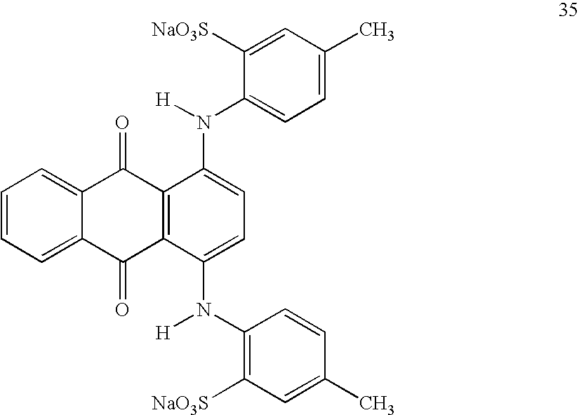
- Aged chicken juice was dropped onto a SCOTT® paper towel. 50 mg of Cl Acid Red 37 (from Aldrich Chemical), structure 36 below and example of an aminoazo dye, was dissolved in 10 ml deionized water. This dye solution was dropped onto the aged chicken juice on the paper towel. No color changes were observed. As a control, 100 mg of Reichardt's dye was suspended in 10 ml acetonitrile. This suspension was dropped onto the aged chicken juice and immediately decolorized.

- Aged chicken juice was dropped onto a SCOTT® paper towel. 50 mg of Cl Acid Yellow 23 (also known as the food colorant tartrazine) (from Aldrich Chemical), structure 37 below, and example of a phenylpyrazolone dye, was dissolved in 10 ml deionized water. This dye solution was dropped onto the aged chicken juice on the paper towel. No color changes were observed. As a control, 100 mg of Reichardt's dye was suspended in 10 ml acetonitrile. This suspension was dropped onto the aged chicken juice and immediately decolorized.

- Aged chicken juice was dropped onto a SCOTT® paper towel. Cl Acid Red 52 (sulforhodamine B), structure 38 below, an example of a xanthene dye, solution in water was dropped onto the aged chicken juice on the paper towel. No color changes were observed. As a control, 100 mg of Reichardt's dye was suspended in 10 ml acetonitrile. This suspension was dropped onto the aged chicken juice and immediately decolorized.

- Aged chicken juice was dropped onto a SCOTT® paper towel. 30 mg of Cl Acid Blue 74 (also known as Indigo Carmine), structure 39 below, (from Aldrich Chemical), as an example of an indigoid dye, was dissolved in 10 ml deionized water. This dye solution was dropped onto the aged chicken juice on the paper towel. No color changes were observed. As a control, 100 mg of Reichardt's dye was suspended in 10 ml acetonitrile. This suspension was dropped onto the aged chicken juice and immediately decolorized.

- As will be appreciated by those skilled in the art, changes and variations to the invention are considered to be within the ability of those skilled in the art. Examples of such changes are contained in the patents identified above, each of which is incorporated herein by reference in its entirety to the extent it is consistent with this specification. Such changes and variations are intended by the inventors to be within the scope of the invention. It is also to be understood that the scope of the present invention is not to be interpreted as limited to the specific embodiments disclosed herein, but only in accordance with the appended claims when read in light of the foregoing disclosure.
Claims (32)


Priority Applications (7)
| Application Number | Priority Date | Filing Date | Title |
|---|---|---|---|
| US11/117,651 US7399608B2 (en) | 2003-12-16 | 2005-04-28 | Microbial detection and quantification |
| KR1020077013575A KR101225911B1 (en) | 2004-12-16 | 2005-10-24 | Microbial detection and quantification |
| CN2005800428210A CN101080497B (en) | 2003-12-16 | 2005-10-24 | Microbial detection and quantification |
| MX2007007122A MX2007007122A (en) | 2004-12-16 | 2005-10-24 | Microbial detection and quantification. |
| CN2010105288572A CN102121056B (en) | 2004-12-16 | 2005-10-24 | Microbial detection and quantification |
| PCT/US2005/038416 WO2006065350A2 (en) | 2004-12-16 | 2005-10-24 | Microbial detection and quantification |
| EP20050851252 EP1828402B1 (en) | 2004-12-16 | 2005-10-24 | Microbial detection and quantification |
Applications Claiming Priority (3)
| Application Number | Priority Date | Filing Date | Title |
|---|---|---|---|
| US10/737,574 US7282349B2 (en) | 2003-12-16 | 2003-12-16 | Solvatochromatic bacterial detection |
| PCT/US2004/042461 WO2005059162A2 (en) | 2003-12-16 | 2004-12-16 | Microbial detection and quantification |
| US11/117,651 US7399608B2 (en) | 2003-12-16 | 2005-04-28 | Microbial detection and quantification |
Related Parent Applications (1)
| Application Number | Title | Priority Date | Filing Date |
|---|---|---|---|
| PCT/US2004/042461 Continuation-In-Part WO2005059162A2 (en) | 2003-12-16 | 2004-12-16 | Microbial detection and quantification |
Publications (2)
| Publication Number | Publication Date |
|---|---|
| US20060134728A1 true US20060134728A1 (en) | 2006-06-22 |
| US7399608B2 US7399608B2 (en) | 2008-07-15 |
Family
ID=36577188
Family Applications (1)
| Application Number | Title | Priority Date | Filing Date |
|---|---|---|---|
| US11/117,651 Expired - Fee Related US7399608B2 (en) | 2003-12-16 | 2005-04-28 | Microbial detection and quantification |
Country Status (3)
| Country | Link |
|---|---|
| US (1) | US7399608B2 (en) |
| EP (1) | EP1828402B1 (en) |
| WO (1) | WO2006065350A2 (en) |
Cited By (38)
| Publication number | Priority date | Publication date | Assignee | Title |
|---|---|---|---|---|
| US20050130253A1 (en) * | 2003-12-16 | 2005-06-16 | Kimberly-Clark Worldwide, Inc. | Solvatochromatic bacterial detection |
| US20060134613A1 (en) * | 2004-12-16 | 2006-06-22 | Kimberly-Clark Worldwide, Inc. | Detection of microbe contamination on elastomeric articles |
| US20070140971A1 (en) * | 2005-12-15 | 2007-06-21 | Macdonald John G | Method for screening for bacterial conjunctivitis |
| US20070293818A1 (en) * | 2006-05-08 | 2007-12-20 | Becton, Dickinson & Company | Vascular access device cleaning status indication |
| US20080057533A1 (en) * | 2006-08-31 | 2008-03-06 | Kimberly-Clark Worldwide, Inc. | Array for rapid detection of a microorganism |
| US20080057534A1 (en) * | 2006-08-31 | 2008-03-06 | Kimberly-Clark Worldwide, Inc. | Microbe-sensitive indicators and use of the same |
| US20080057532A1 (en) * | 2006-08-31 | 2008-03-06 | Stephanie Martin | Method for detecting Candida on skin |
| US20080060550A1 (en) * | 2006-09-12 | 2008-03-13 | Macdonald Gavin | Color changing skin sealant with co-acid trigger |
| US20080063615A1 (en) * | 2006-09-12 | 2008-03-13 | Macdonald John Gavin | Color changing skin sealant |
| WO2008051892A2 (en) * | 2006-10-23 | 2008-05-02 | The Clorox Company | One-handed method of cleaning surfaces |
| US20080145316A1 (en) * | 2006-12-14 | 2008-06-19 | Macdonald John Gavin | Skin coating with microbial indicator |
| US20090018047A1 (en) * | 2007-07-12 | 2009-01-15 | Kimberly-Clark Worldwide, Inc. | Foaming hand sponge for bodily cleansing with color change indicator |
| US20090018048A1 (en) * | 2007-07-12 | 2009-01-15 | Kimberly-Clark Worldwide, Inc. | Foaming hand sponge with color change indicator |
| US20090024096A1 (en) * | 2007-07-20 | 2009-01-22 | Baxter International Inc. | Immobilization of dyes and antimicrobial agents on a medical device |
| US20090042757A1 (en) * | 2005-03-30 | 2009-02-12 | Carling Philip C | Monitoring Cleaning of Surfaces |
| US20090112295A1 (en) * | 2007-10-30 | 2009-04-30 | Searete Llc, A Limited Liability Corporation Of The State Of Delaware | Devices and systems that deliver nitric oxide |
| US20090110958A1 (en) * | 2007-10-30 | 2009-04-30 | Searete Llc, A Limited Liability Corporation Of The State Of Delaware | Systems and devices that utilize photolyzable nitric oxide donors |
| US20090108777A1 (en) * | 2007-10-30 | 2009-04-30 | Searete Llc, A Limited Liability Corporation Of The State Of Delaware | Devices and systems that deliver nitric oxide |
| US20090110604A1 (en) * | 2007-10-30 | 2009-04-30 | Searete Llc, A Limited Liability Corporation Of The State Of Delaware | Substrates for nitric oxide releasing devices |
| US20090110712A1 (en) * | 2007-10-30 | 2009-04-30 | Searete Llc, A Limited Liability Corporation Of The State Of Delaware | Methods and systems for use of photolyzable nitric oxide donors |
| US20090123569A1 (en) * | 2007-11-08 | 2009-05-14 | Macdonald John Gavin | Coverage indicating technology for skin sealants using tannates |
| US20090288366A1 (en) * | 2008-05-23 | 2009-11-26 | Phillip Andrew Schorr | Vacuum packaged products and methods for making same |
| US20090325221A1 (en) * | 2008-06-30 | 2009-12-31 | Kimberly-Clark Worldwide, Inc. | Temporary Tattoo Decals for Detecting the Presence of an Analyte |
| US20100197817A1 (en) * | 2007-07-20 | 2010-08-05 | Baxter International Inc. | Antimicrobial housing and cover for a medical device |
| US20100196636A1 (en) * | 2009-01-26 | 2010-08-05 | Indicator Systems International, Inc. | Indicators for detecting the presence of metabolic byproducts from microorganisms |
| EP2224965A1 (en) * | 2007-12-13 | 2010-09-08 | Kimberly-Clark Worldwide, Inc. | Self-indicating wipe for removing bacteria from a surface |
| WO2010107979A1 (en) * | 2009-03-19 | 2010-09-23 | Tangidyne Corporation | Detection device and method for detecting analyte |
| US7897399B2 (en) | 2007-10-30 | 2011-03-01 | The Invention Science Fund I, Llc | Nitric oxide sensors and systems |
| US7975699B2 (en) | 2007-10-30 | 2011-07-12 | The Invention Science Fund I, Llc | Condoms configured to facilitate release of nitric oxide |
| US20110190604A1 (en) * | 2006-12-22 | 2011-08-04 | Hyde Roderick A | Nitric oxide sensors and systems |
| US20120028879A1 (en) * | 2009-03-31 | 2012-02-02 | National University Corporation Hamamatsu University School Of Medicine | Highly sensitive detection method for highly virulent oral cavity bacteria |
| EP2428580A1 (en) * | 2009-05-07 | 2012-03-14 | Universidad De Zaragoza | Smart packaging for detecting microorganisms |
| US10080823B2 (en) | 2007-10-30 | 2018-09-25 | Gearbox Llc | Substrates for nitric oxide releasing devices |
| USRE47452E1 (en) * | 2007-07-20 | 2019-06-25 | Baxter International Inc. | Antimicrobial housing and cover for a medical device |
| CN111445444A (en) * | 2020-03-11 | 2020-07-24 | 中南大学 | Molten iron flow velocity detection method based on polarization characteristics |
| WO2020183072A1 (en) * | 2019-03-14 | 2020-09-17 | Hitachi High-Tech Corporation | Method for testing sensitivity to chemical agents |
| US11739362B2 (en) | 2017-04-04 | 2023-08-29 | Fresh Check Ltd. | Colour changing compositions |
| US11814668B2 (en) | 2020-02-14 | 2023-11-14 | Phigenics, Llc | Detection of Mycobacterium on growth media |
Families Citing this family (12)
| Publication number | Priority date | Publication date | Assignee | Title |
|---|---|---|---|---|
| JP4912323B2 (en) * | 2005-12-14 | 2012-04-11 | 日本製紙株式会社 | Coated paper for printing |
| WO2007142302A1 (en) * | 2006-06-07 | 2007-12-13 | Nippon Paper Industries Co., Ltd. | Cast-coated paper |
| US20090111088A1 (en) * | 2007-08-30 | 2009-04-30 | Kimberly-Clark Worldwide, Inc. | Rapid assessment of upper respiratory conditions |
| US8980332B2 (en) | 2007-10-30 | 2015-03-17 | The Invention Science Fund I, Llc | Methods and systems for use of photolyzable nitric oxide donors |
| US20090142275A1 (en) * | 2007-11-29 | 2009-06-04 | Kimberly-Clark Worldwide, Inc. | Wound Suture Capable of Identifying the Presence of Bacteria |
| US8247220B2 (en) * | 2008-02-01 | 2012-08-21 | Kimberly-Clark Worldwide, Inc. | Optical indicator for detecting bacterial pathogens |
| AT509355B1 (en) | 2010-02-10 | 2012-04-15 | Univ Graz Tech | TEST ARRANGEMENT |
| US9018434B2 (en) | 2010-08-06 | 2015-04-28 | Kimberly-Clark Worldwide, Inc. | Absorbent articles with intricate graphics |
| USD656852S1 (en) | 2010-08-06 | 2012-04-03 | Kimberly-Clark Worldwide, Inc. | Wetness indicator |
| US9034593B2 (en) | 2010-11-22 | 2015-05-19 | Kimberly-Clark Worldwide, Inc. | Vaginal indicator to detect biomarkers of good health |
| JP7421504B2 (en) | 2018-06-01 | 2024-01-24 | ビオラ・セラピューティクス・インコーポレイテッド | Devices and systems for gastrointestinal microbiome detection and manipulation |
| RU210196U1 (en) * | 2020-07-27 | 2022-03-31 | Федеральное бюджетное учреждение науки "Санкт-Петербургский научно-исследовательский институт эпидемиологии и микробиологии им. Пастера Федеральной службы по надзору в сфере защиты прав потребителей и благополучия человека" (ФБУН НИИ эпидемиологии и микробиологии имени Пастера) | Disk for preparing liquid culture media containing pH or redox indicators |
Citations (45)
| Publication number | Priority date | Publication date | Assignee | Title |
|---|---|---|---|---|
| US3338992A (en) * | 1959-12-15 | 1967-08-29 | Du Pont | Process for forming non-woven filamentary structures from fiber-forming synthetic organic polymers |
| US3341394A (en) * | 1966-12-21 | 1967-09-12 | Du Pont | Sheets of randomly distributed continuous filaments |
| US3502763A (en) * | 1962-02-03 | 1970-03-24 | Freudenberg Carl Kg | Process of producing non-woven fabric fleece |
| US3542615A (en) * | 1967-06-16 | 1970-11-24 | Monsanto Co | Process for producing a nylon non-woven fabric |
| US3692618A (en) * | 1969-10-08 | 1972-09-19 | Metallgesellschaft Ag | Continuous filament nonwoven web |
| US3705032A (en) * | 1968-08-20 | 1972-12-05 | Fuji Photo Film Co Ltd | Electrophotographic materials |
| US3802817A (en) * | 1969-10-01 | 1974-04-09 | Asahi Chemical Ind | Apparatus for producing non-woven fleeces |
| US3849241A (en) * | 1968-12-23 | 1974-11-19 | Exxon Research Engineering Co | Non-woven mats by melt blowing |
| US4100324A (en) * | 1974-03-26 | 1978-07-11 | Kimberly-Clark Corporation | Nonwoven fabric and method of producing same |
| US4310928A (en) * | 1979-07-30 | 1982-01-19 | American Hospital Supply Corporation | Surgeon's glove and talc free process for forming same |
| US4340563A (en) * | 1980-05-05 | 1982-07-20 | Kimberly-Clark Corporation | Method for forming nonwoven webs |
| US4340395A (en) * | 1979-11-13 | 1982-07-20 | Miles Laboratories, Inc. | Stabilization of benzidine-type indicators with various enhancers |
| US4375448A (en) * | 1979-12-21 | 1983-03-01 | Kimberly-Clark Corporation | Method of forming a web of air-laid dry fibers |
| US4494278A (en) * | 1977-11-08 | 1985-01-22 | Karl Kristian Kobs Kroyer | Apparatus for the production of a fibrous web |
| US4511488A (en) * | 1983-12-05 | 1985-04-16 | Penetone Corporation | D-Limonene based aqueous cleaning compositions |
| US4556636A (en) * | 1983-06-09 | 1985-12-03 | Eastman Kodak Company | Composition, analytical element and method for the detection of bacteria |
| US4640810A (en) * | 1984-06-12 | 1987-02-03 | Scan Web Of North America, Inc. | System for producing an air laid web |
| US4677076A (en) * | 1980-11-21 | 1987-06-30 | Heinz Langhals | Process for determining water in samples containing water |
| US4775582A (en) * | 1986-08-15 | 1988-10-04 | Kimberly-Clark Corporation | Uniformly moist wipes |
| US4780422A (en) * | 1986-06-27 | 1988-10-25 | Tokuyama Soda Kabushiki Kaisha | Dyed inorganic composite particles and process for production thereof |
| US4818464A (en) * | 1984-08-30 | 1989-04-04 | Kimberly-Clark Corporation | Extrusion process using a central air jet |
| US4833003A (en) * | 1986-08-15 | 1989-05-23 | Kimberly-Clark Corporation | Uniformly moist abrasive wipes |
| US4853281A (en) * | 1986-08-15 | 1989-08-01 | Kimberly-Clark Corporation | Uniformly moist wipes |
| US5036000A (en) * | 1986-12-16 | 1991-07-30 | Enzymatics, Inc. | Threshold color control system |
| US5112900A (en) * | 1990-11-28 | 1992-05-12 | Tactyl Technologies, Inc. | Elastomeric triblock copolymer compositions and articles made therewith |
| US5407715A (en) * | 1990-11-28 | 1995-04-18 | Tactyl Technologies, Inc. | Elastomeric triblock copolymer compositions and articles made therewith |
| US5464739A (en) * | 1994-08-22 | 1995-11-07 | Bayer Corporation | Composition method for determining the presence of leukocyte cells, esterase or protease in a test sample |
| US5468469A (en) * | 1992-08-11 | 1995-11-21 | The United States Of America As Represented By The Department Of Health And Human Services | Methods of using azo dyes and their derivatives |
| US5527171A (en) * | 1993-03-09 | 1996-06-18 | Niro Separation A/S | Apparatus for depositing fibers |
| US5534416A (en) * | 1993-04-13 | 1996-07-09 | Molecular Probes, Inc. | Fluorescent viability assay using cyclic-substituted unsymmetrical cyanine dyes |
| US5742943A (en) * | 1996-06-28 | 1998-04-28 | Johnson & Johnson Medical, Inc. | Slip-coated elastomeric flexible articles and their method of manufacture |
| US5744321A (en) * | 1997-02-21 | 1998-04-28 | Gem Biomedical, Inc. | Detection of fish spoilage by colorimetry |
| US5792531A (en) * | 1996-02-20 | 1998-08-11 | Tactyl Technologies, Inc. | Readily donned, powder free elastomeric article |
| US5900452A (en) * | 1996-08-12 | 1999-05-04 | Tactyl Technologies, Inc. | S-EB-S block copolymer/oil aqueous dispersion and its use in forming articles |
| US6090541A (en) * | 1997-04-18 | 2000-07-18 | 3M Innovative Properties Company | Method and device for detecting bacteriophage using contrast-coloring and precipitable dyes |
| US6168655B1 (en) * | 1995-11-28 | 2001-01-02 | Kimberly-Clark Worldwide, Inc. | Colorant stabilizers |
| US6306514B1 (en) * | 1996-12-31 | 2001-10-23 | Ansell Healthcare Products Inc. | Slip-coated elastomeric flexible articles and their method of manufacture |
| US6362006B1 (en) * | 2000-03-13 | 2002-03-26 | General Electric Company | Rapid parallel determination of non-volatile analytes in complex combinatorial samples |
| US6368558B1 (en) * | 2000-03-21 | 2002-04-09 | The Board Of Trustees Of The University Of Illinois | Colorimetric artificial nose having an array of dyes and method for artificial olfaction |
| US6383815B1 (en) * | 2001-04-04 | 2002-05-07 | General Electric Company | Devices and methods for measurements of barrier properties of coating arrays |
| US6524846B1 (en) * | 2001-11-05 | 2003-02-25 | William L. Robinson, Jr. | Biological toxin detection system for mailed materials |
| US20030119202A1 (en) * | 2001-12-24 | 2003-06-26 | Kimberly-Clark Worldwide, Inc. | Reading device, method, and system for conducting lateral flow assays |
| US20030143112A1 (en) * | 2000-03-21 | 2003-07-31 | Board Of Trustees Of The University Of Illinois | Colorimetric artificial nose having an array of dyes and method for artificial olfaction |
| US20050130253A1 (en) * | 2003-12-16 | 2005-06-16 | Kimberly-Clark Worldwide, Inc. | Solvatochromatic bacterial detection |
| US20060134613A1 (en) * | 2004-12-16 | 2006-06-22 | Kimberly-Clark Worldwide, Inc. | Detection of microbe contamination on elastomeric articles |
Family Cites Families (11)
| Publication number | Priority date | Publication date | Assignee | Title |
|---|---|---|---|---|
| DE1598092A1 (en) | 1966-03-29 | 1970-04-23 | Boehringer Mannheim Gmbh | Methods and diagnostic means for the detection of bacteria |
| GB2178847A (en) | 1985-08-07 | 1987-02-18 | Philips Electronic Associated | Testing for the presence of living organisms at the surface of an object |
| US4859584A (en) | 1986-10-31 | 1989-08-22 | Smithkline Beckman Corporation | Cell growth rate determination by measurement of changes in cyanine dye levels in plasma membranes |
| DE69213826T2 (en) | 1991-06-06 | 1997-01-30 | Bayer Ag | Test strips with merocyanine and nitro or nitroso substituted polyhalogenated phenolsulfonphthaleins as protein indicators |
| US5703364A (en) | 1996-02-15 | 1997-12-30 | Futrex, Inc. | Method and apparatus for near-infrared quantitative analysis |
| US6225083B1 (en) | 1996-10-01 | 2001-05-01 | Smithkline Beecham Corporation | FtsL from Streptococcus pneumoniae |
| JPH1183849A (en) * | 1997-09-12 | 1999-03-26 | Toa Medical Electronics Co Ltd | Analyzing method for material component in urine and reagent for its analysis |
| GB0025245D0 (en) | 2000-10-14 | 2000-11-29 | Lee Helen | Multiple target detection |
| IL143810A (en) | 2001-06-18 | 2008-06-05 | Alexander Schoenfeld | Prophylactic article useful for both protection and diagnosis and method for use and production thereof |
| WO2003065019A1 (en) | 2002-01-25 | 2003-08-07 | Illumina, Inc. | Sensor arrays for detecting analytes in fluids |
| AU2003304409A1 (en) | 2002-06-07 | 2005-03-07 | President And Fellows Of Harvard College | Evaluating protein signatures |
-
2005
- 2005-04-28 US US11/117,651 patent/US7399608B2/en not_active Expired - Fee Related
- 2005-10-24 WO PCT/US2005/038416 patent/WO2006065350A2/en active Application Filing
- 2005-10-24 EP EP20050851252 patent/EP1828402B1/en not_active Expired - Fee Related
Patent Citations (47)
| Publication number | Priority date | Publication date | Assignee | Title |
|---|---|---|---|---|
| US3338992A (en) * | 1959-12-15 | 1967-08-29 | Du Pont | Process for forming non-woven filamentary structures from fiber-forming synthetic organic polymers |
| US3502763A (en) * | 1962-02-03 | 1970-03-24 | Freudenberg Carl Kg | Process of producing non-woven fabric fleece |
| US3341394A (en) * | 1966-12-21 | 1967-09-12 | Du Pont | Sheets of randomly distributed continuous filaments |
| US3542615A (en) * | 1967-06-16 | 1970-11-24 | Monsanto Co | Process for producing a nylon non-woven fabric |
| US3705032A (en) * | 1968-08-20 | 1972-12-05 | Fuji Photo Film Co Ltd | Electrophotographic materials |
| US3849241A (en) * | 1968-12-23 | 1974-11-19 | Exxon Research Engineering Co | Non-woven mats by melt blowing |
| US3802817A (en) * | 1969-10-01 | 1974-04-09 | Asahi Chemical Ind | Apparatus for producing non-woven fleeces |
| US3692618A (en) * | 1969-10-08 | 1972-09-19 | Metallgesellschaft Ag | Continuous filament nonwoven web |
| US4100324A (en) * | 1974-03-26 | 1978-07-11 | Kimberly-Clark Corporation | Nonwoven fabric and method of producing same |
| US4494278A (en) * | 1977-11-08 | 1985-01-22 | Karl Kristian Kobs Kroyer | Apparatus for the production of a fibrous web |
| US4310928A (en) * | 1979-07-30 | 1982-01-19 | American Hospital Supply Corporation | Surgeon's glove and talc free process for forming same |
| US4340395A (en) * | 1979-11-13 | 1982-07-20 | Miles Laboratories, Inc. | Stabilization of benzidine-type indicators with various enhancers |
| US4375448A (en) * | 1979-12-21 | 1983-03-01 | Kimberly-Clark Corporation | Method of forming a web of air-laid dry fibers |
| US4340563A (en) * | 1980-05-05 | 1982-07-20 | Kimberly-Clark Corporation | Method for forming nonwoven webs |
| US4677076A (en) * | 1980-11-21 | 1987-06-30 | Heinz Langhals | Process for determining water in samples containing water |
| US4556636A (en) * | 1983-06-09 | 1985-12-03 | Eastman Kodak Company | Composition, analytical element and method for the detection of bacteria |
| US4511488A (en) * | 1983-12-05 | 1985-04-16 | Penetone Corporation | D-Limonene based aqueous cleaning compositions |
| US4511488B1 (en) * | 1983-12-05 | 1990-09-11 | Penetone Corp | |
| US4640810A (en) * | 1984-06-12 | 1987-02-03 | Scan Web Of North America, Inc. | System for producing an air laid web |
| US4818464A (en) * | 1984-08-30 | 1989-04-04 | Kimberly-Clark Corporation | Extrusion process using a central air jet |
| US4780422A (en) * | 1986-06-27 | 1988-10-25 | Tokuyama Soda Kabushiki Kaisha | Dyed inorganic composite particles and process for production thereof |
| US4853281A (en) * | 1986-08-15 | 1989-08-01 | Kimberly-Clark Corporation | Uniformly moist wipes |
| US4833003A (en) * | 1986-08-15 | 1989-05-23 | Kimberly-Clark Corporation | Uniformly moist abrasive wipes |
| US4775582A (en) * | 1986-08-15 | 1988-10-04 | Kimberly-Clark Corporation | Uniformly moist wipes |
| US5036000A (en) * | 1986-12-16 | 1991-07-30 | Enzymatics, Inc. | Threshold color control system |
| US5112900A (en) * | 1990-11-28 | 1992-05-12 | Tactyl Technologies, Inc. | Elastomeric triblock copolymer compositions and articles made therewith |
| US5407715A (en) * | 1990-11-28 | 1995-04-18 | Tactyl Technologies, Inc. | Elastomeric triblock copolymer compositions and articles made therewith |
| US5468469A (en) * | 1992-08-11 | 1995-11-21 | The United States Of America As Represented By The Department Of Health And Human Services | Methods of using azo dyes and their derivatives |
| US5527171A (en) * | 1993-03-09 | 1996-06-18 | Niro Separation A/S | Apparatus for depositing fibers |
| US5534416A (en) * | 1993-04-13 | 1996-07-09 | Molecular Probes, Inc. | Fluorescent viability assay using cyclic-substituted unsymmetrical cyanine dyes |
| US5464739A (en) * | 1994-08-22 | 1995-11-07 | Bayer Corporation | Composition method for determining the presence of leukocyte cells, esterase or protease in a test sample |
| US6168655B1 (en) * | 1995-11-28 | 2001-01-02 | Kimberly-Clark Worldwide, Inc. | Colorant stabilizers |
| US5792531A (en) * | 1996-02-20 | 1998-08-11 | Tactyl Technologies, Inc. | Readily donned, powder free elastomeric article |
| US5742943A (en) * | 1996-06-28 | 1998-04-28 | Johnson & Johnson Medical, Inc. | Slip-coated elastomeric flexible articles and their method of manufacture |
| US5900452A (en) * | 1996-08-12 | 1999-05-04 | Tactyl Technologies, Inc. | S-EB-S block copolymer/oil aqueous dispersion and its use in forming articles |
| US6288159B1 (en) * | 1996-08-12 | 2001-09-11 | Tactyl Technologies, Inc. | S-EB-S block copolymer/oil aqueous dispersion and its use in forming articles |
| US6306514B1 (en) * | 1996-12-31 | 2001-10-23 | Ansell Healthcare Products Inc. | Slip-coated elastomeric flexible articles and their method of manufacture |
| US5744321A (en) * | 1997-02-21 | 1998-04-28 | Gem Biomedical, Inc. | Detection of fish spoilage by colorimetry |
| US6090541A (en) * | 1997-04-18 | 2000-07-18 | 3M Innovative Properties Company | Method and device for detecting bacteriophage using contrast-coloring and precipitable dyes |
| US6362006B1 (en) * | 2000-03-13 | 2002-03-26 | General Electric Company | Rapid parallel determination of non-volatile analytes in complex combinatorial samples |
| US6368558B1 (en) * | 2000-03-21 | 2002-04-09 | The Board Of Trustees Of The University Of Illinois | Colorimetric artificial nose having an array of dyes and method for artificial olfaction |
| US20030143112A1 (en) * | 2000-03-21 | 2003-07-31 | Board Of Trustees Of The University Of Illinois | Colorimetric artificial nose having an array of dyes and method for artificial olfaction |
| US6383815B1 (en) * | 2001-04-04 | 2002-05-07 | General Electric Company | Devices and methods for measurements of barrier properties of coating arrays |
| US6524846B1 (en) * | 2001-11-05 | 2003-02-25 | William L. Robinson, Jr. | Biological toxin detection system for mailed materials |
| US20030119202A1 (en) * | 2001-12-24 | 2003-06-26 | Kimberly-Clark Worldwide, Inc. | Reading device, method, and system for conducting lateral flow assays |
| US20050130253A1 (en) * | 2003-12-16 | 2005-06-16 | Kimberly-Clark Worldwide, Inc. | Solvatochromatic bacterial detection |
| US20060134613A1 (en) * | 2004-12-16 | 2006-06-22 | Kimberly-Clark Worldwide, Inc. | Detection of microbe contamination on elastomeric articles |
Cited By (72)
| Publication number | Priority date | Publication date | Assignee | Title |
|---|---|---|---|---|
| US7282349B2 (en) | 2003-12-16 | 2007-10-16 | Kimberly-Clark Worldwide, Inc. | Solvatochromatic bacterial detection |
| US20050130253A1 (en) * | 2003-12-16 | 2005-06-16 | Kimberly-Clark Worldwide, Inc. | Solvatochromatic bacterial detection |
| US20060134613A1 (en) * | 2004-12-16 | 2006-06-22 | Kimberly-Clark Worldwide, Inc. | Detection of microbe contamination on elastomeric articles |
| US7300770B2 (en) | 2004-12-16 | 2007-11-27 | Kimberly-Clark Worldwide, Inc. | Detection of microbe contamination on elastomeric articles |
| US8435933B2 (en) | 2005-03-30 | 2013-05-07 | Kleancheck Systems, Llc | Monitoring cleaning of surfaces |
| US9624456B2 (en) | 2005-03-30 | 2017-04-18 | Kleancheck Systems, Llc | Monitoring cleaning of surfaces |
| US9458417B2 (en) | 2005-03-30 | 2016-10-04 | Kleancheck Systems, Llc | Monitoring cleaning of surfaces |
| US11078446B2 (en) | 2005-03-30 | 2021-08-03 | Kleancheck Systems, Llc | Monitoring cleaning of surfaces |
| US8084410B2 (en) * | 2005-03-30 | 2011-12-27 | Kleancheck Systems, Llc | Monitoring cleaning of surfaces |
| US20090042757A1 (en) * | 2005-03-30 | 2009-02-12 | Carling Philip C | Monitoring Cleaning of Surfaces |
| US20070140971A1 (en) * | 2005-12-15 | 2007-06-21 | Macdonald John G | Method for screening for bacterial conjunctivitis |
| US7727513B2 (en) * | 2005-12-15 | 2010-06-01 | Kimberly-Clark Worldwide, Inc. | Method for screening for bacterial conjunctivitis |
| US20070293818A1 (en) * | 2006-05-08 | 2007-12-20 | Becton, Dickinson & Company | Vascular access device cleaning status indication |
| US8137303B2 (en) * | 2006-05-08 | 2012-03-20 | Becton, Dickinson And Company | Vascular access device cleaning status indication |
| US8617874B2 (en) | 2006-08-31 | 2013-12-31 | Kimberly-Clark Worldwide, Inc. | Array for rapid detection of a microorganism |
| US7763442B2 (en) | 2006-08-31 | 2010-07-27 | Kimberly-Clark Worldwide, Inc. | Method for detecting candida on skin |
| US8361742B2 (en) | 2006-08-31 | 2013-01-29 | Kimberly-Clark Worldwide, Inc. | Method for detecting Candida on skin |
| US20090221061A1 (en) * | 2006-08-31 | 2009-09-03 | Kimberly-Clark Worldwide, Inc. | Array for Rapid Detection of a Microorganism |
| US20080057532A1 (en) * | 2006-08-31 | 2008-03-06 | Stephanie Martin | Method for detecting Candida on skin |
| US20080057534A1 (en) * | 2006-08-31 | 2008-03-06 | Kimberly-Clark Worldwide, Inc. | Microbe-sensitive indicators and use of the same |
| US20080057533A1 (en) * | 2006-08-31 | 2008-03-06 | Kimberly-Clark Worldwide, Inc. | Array for rapid detection of a microorganism |
| US7531319B2 (en) | 2006-08-31 | 2009-05-12 | Kimberly-Clark Worldwide, Inc. | Array for rapid detection of a microorganism |
| US20080060550A1 (en) * | 2006-09-12 | 2008-03-13 | Macdonald Gavin | Color changing skin sealant with co-acid trigger |
| US20080063615A1 (en) * | 2006-09-12 | 2008-03-13 | Macdonald John Gavin | Color changing skin sealant |
| WO2008051892A2 (en) * | 2006-10-23 | 2008-05-02 | The Clorox Company | One-handed method of cleaning surfaces |
| WO2008051892A3 (en) * | 2006-10-23 | 2008-12-11 | Clorox Co | One-handed method of cleaning surfaces |
| US20080145316A1 (en) * | 2006-12-14 | 2008-06-19 | Macdonald John Gavin | Skin coating with microbial indicator |
| US20110190604A1 (en) * | 2006-12-22 | 2011-08-04 | Hyde Roderick A | Nitric oxide sensors and systems |
| US20090018048A1 (en) * | 2007-07-12 | 2009-01-15 | Kimberly-Clark Worldwide, Inc. | Foaming hand sponge with color change indicator |
| US20090018047A1 (en) * | 2007-07-12 | 2009-01-15 | Kimberly-Clark Worldwide, Inc. | Foaming hand sponge for bodily cleansing with color change indicator |
| US8440606B2 (en) | 2007-07-12 | 2013-05-14 | Kimberly-Clark Worldwide, Inc. | Foaming hand sponge for bodily cleansing with color change indicator |
| US8343908B2 (en) | 2007-07-12 | 2013-01-01 | Kimberly-Clark Worldwide, Inc. | Foaming hand sponge with color change indicator |
| US20090024096A1 (en) * | 2007-07-20 | 2009-01-22 | Baxter International Inc. | Immobilization of dyes and antimicrobial agents on a medical device |
| CN101801429A (en) * | 2007-07-20 | 2010-08-11 | 巴克斯特国际公司 | The immobilization on medical treatment device of dyestuff and antimicrobial |
| US20100197817A1 (en) * | 2007-07-20 | 2010-08-05 | Baxter International Inc. | Antimicrobial housing and cover for a medical device |
| US9125973B2 (en) * | 2007-07-20 | 2015-09-08 | Baxter International Inc. | Antimicrobial housing and cover for a medical device |
| US9574060B2 (en) * | 2007-07-20 | 2017-02-21 | Baxter International Inc. | Antimicrobial housing and cover for a medical device |
| USRE47452E1 (en) * | 2007-07-20 | 2019-06-25 | Baxter International Inc. | Antimicrobial housing and cover for a medical device |
| US20150322229A1 (en) * | 2007-07-20 | 2015-11-12 | Baxter International Inc. | Antimicrobial housing and cover for a medical device |
| US20090112295A1 (en) * | 2007-10-30 | 2009-04-30 | Searete Llc, A Limited Liability Corporation Of The State Of Delaware | Devices and systems that deliver nitric oxide |
| US10080823B2 (en) | 2007-10-30 | 2018-09-25 | Gearbox Llc | Substrates for nitric oxide releasing devices |
| US7897399B2 (en) | 2007-10-30 | 2011-03-01 | The Invention Science Fund I, Llc | Nitric oxide sensors and systems |
| US20090110958A1 (en) * | 2007-10-30 | 2009-04-30 | Searete Llc, A Limited Liability Corporation Of The State Of Delaware | Systems and devices that utilize photolyzable nitric oxide donors |
| US20090108777A1 (en) * | 2007-10-30 | 2009-04-30 | Searete Llc, A Limited Liability Corporation Of The State Of Delaware | Devices and systems that deliver nitric oxide |
| US8642093B2 (en) | 2007-10-30 | 2014-02-04 | The Invention Science Fund I, Llc | Methods and systems for use of photolyzable nitric oxide donors |
| US8221690B2 (en) | 2007-10-30 | 2012-07-17 | The Invention Science Fund I, Llc | Systems and devices that utilize photolyzable nitric oxide donors |
| US20090110712A1 (en) * | 2007-10-30 | 2009-04-30 | Searete Llc, A Limited Liability Corporation Of The State Of Delaware | Methods and systems for use of photolyzable nitric oxide donors |
| US20090110604A1 (en) * | 2007-10-30 | 2009-04-30 | Searete Llc, A Limited Liability Corporation Of The State Of Delaware | Substrates for nitric oxide releasing devices |
| US7975699B2 (en) | 2007-10-30 | 2011-07-12 | The Invention Science Fund I, Llc | Condoms configured to facilitate release of nitric oxide |
| US7862598B2 (en) | 2007-10-30 | 2011-01-04 | The Invention Science Fund I, Llc | Devices and systems that deliver nitric oxide |
| US8349262B2 (en) | 2007-10-30 | 2013-01-08 | The Invention Science Fund I, Llc | Nitric oxide permeable housings |
| US7846400B2 (en) | 2007-10-30 | 2010-12-07 | The Invention Science Fund I, Llc | Substrates for nitric oxide releasing devices |
| US8877508B2 (en) | 2007-10-30 | 2014-11-04 | The Invention Science Fund I, Llc | Devices and systems that deliver nitric oxide |
| US20090123569A1 (en) * | 2007-11-08 | 2009-05-14 | Macdonald John Gavin | Coverage indicating technology for skin sealants using tannates |
| EP2224965A4 (en) * | 2007-12-13 | 2011-01-05 | Kimberly Clark Co | Self-indicating wipe for removing bacteria from a surface |
| EP2224965A1 (en) * | 2007-12-13 | 2010-09-08 | Kimberly-Clark Worldwide, Inc. | Self-indicating wipe for removing bacteria from a surface |
| AU2008334327B2 (en) * | 2007-12-13 | 2013-03-28 | Kimberly-Clark Worldwide, Inc. | Self-indicating wipe for removing bacteria from a surface |
| US8323562B2 (en) | 2008-05-23 | 2012-12-04 | Kimberly-Clark Worldwide, Inc. | Vacuum packaged products and methods for making same |
| US20090288366A1 (en) * | 2008-05-23 | 2009-11-26 | Phillip Andrew Schorr | Vacuum packaged products and methods for making same |
| US20090325221A1 (en) * | 2008-06-30 | 2009-12-31 | Kimberly-Clark Worldwide, Inc. | Temporary Tattoo Decals for Detecting the Presence of an Analyte |
| US20100196636A1 (en) * | 2009-01-26 | 2010-08-05 | Indicator Systems International, Inc. | Indicators for detecting the presence of metabolic byproducts from microorganisms |
| US8425996B2 (en) * | 2009-01-26 | 2013-04-23 | Indicator Systems International, Inc. | Indicators for detecting the presence of metabolic byproducts from microorganisms |
| WO2010107979A1 (en) * | 2009-03-19 | 2010-09-23 | Tangidyne Corporation | Detection device and method for detecting analyte |
| US20120028879A1 (en) * | 2009-03-31 | 2012-02-02 | National University Corporation Hamamatsu University School Of Medicine | Highly sensitive detection method for highly virulent oral cavity bacteria |
| JP2012525832A (en) * | 2009-05-07 | 2012-10-25 | ウニヴェルシダド デ サラゴサ | Smart packaging for microbial detection |
| EP2428580A4 (en) * | 2009-05-07 | 2012-10-10 | Univ Zaragoza | Smart packaging for detecting microorganisms |
| EP2428580A1 (en) * | 2009-05-07 | 2012-03-14 | Universidad De Zaragoza | Smart packaging for detecting microorganisms |
| US8741596B2 (en) | 2009-05-07 | 2014-06-03 | Universidad De Zaragoza | Smart packaging for detecting microorganisms |
| US11739362B2 (en) | 2017-04-04 | 2023-08-29 | Fresh Check Ltd. | Colour changing compositions |
| WO2020183072A1 (en) * | 2019-03-14 | 2020-09-17 | Hitachi High-Tech Corporation | Method for testing sensitivity to chemical agents |
| US11814668B2 (en) | 2020-02-14 | 2023-11-14 | Phigenics, Llc | Detection of Mycobacterium on growth media |
| CN111445444A (en) * | 2020-03-11 | 2020-07-24 | 中南大学 | Molten iron flow velocity detection method based on polarization characteristics |
Also Published As
| Publication number | Publication date |
|---|---|
| EP1828402A2 (en) | 2007-09-05 |
| US7399608B2 (en) | 2008-07-15 |
| EP1828402B1 (en) | 2012-06-13 |
| WO2006065350A2 (en) | 2006-06-22 |
| WO2006065350A3 (en) | 2006-08-10 |
Similar Documents
| Publication | Publication Date | Title |
|---|---|---|
| US7399608B2 (en) | Microbial detection and quantification | |
| US7687245B2 (en) | Microbial detection and quantification | |
| US7300770B2 (en) | Detection of microbe contamination on elastomeric articles | |
| EP2059805B1 (en) | Microbe-sensitive indicators and use of the same | |
| EP1828400B1 (en) | Detection of microbe contamination on elastomeric articles | |
| US7727513B2 (en) | Method for screening for bacterial conjunctivitis | |
| KR101811876B1 (en) | Optical indicator for detecting bacterial pathogens | |
| Griffith | Surface sampling and the detection of contamination | |
| KR20100058514A (en) | Rapid assessment of upper respiratory conditions | |
| CN101080497B (en) | Microbial detection and quantification | |
| KR101225911B1 (en) | Microbial detection and quantification |
Legal Events
| Date | Code | Title | Description |
|---|---|---|---|
| AS | Assignment |
Owner name: KIMBERLY-CLARK WORLDWIDE, INC., WISCONSIN Free format text: ASSIGNMENT OF ASSIGNORS INTEREST;ASSIGNORS:MACDONALD, J. GAVIN;MARTIN, STEPHANIE M.;LYE, JASON;REEL/FRAME:016894/0743;SIGNING DATES FROM 20050727 TO 20050802 |
|
| STCF | Information on status: patent grant |
Free format text: PATENTED CASE |
|
| FPAY | Fee payment |
Year of fee payment: 4 |
|
| AS | Assignment |
Owner name: KIMBERLY-CLARK WORLDWIDE, INC., WISCONSIN Free format text: NAME CHANGE;ASSIGNOR:KIMBERLY-CLARK WORLDWIDE, INC.;REEL/FRAME:034880/0742 Effective date: 20150101 |
|
| FPAY | Fee payment |
Year of fee payment: 8 |
|
| FEPP | Fee payment procedure |
Free format text: MAINTENANCE FEE REMINDER MAILED (ORIGINAL EVENT CODE: REM.); ENTITY STATUS OF PATENT OWNER: LARGE ENTITY |
|
| LAPS | Lapse for failure to pay maintenance fees |
Free format text: PATENT EXPIRED FOR FAILURE TO PAY MAINTENANCE FEES (ORIGINAL EVENT CODE: EXP.); ENTITY STATUS OF PATENT OWNER: LARGE ENTITY |
|
| STCH | Information on status: patent discontinuation |
Free format text: PATENT EXPIRED DUE TO NONPAYMENT OF MAINTENANCE FEES UNDER 37 CFR 1.362 |
|
| FP | Lapsed due to failure to pay maintenance fee |
Effective date: 20200715 |